Run your tongue along your teeth from the front to the back. The front ones have thin, sharp edges; the ones at the back are broad and bumpy. In the egg-white experiment you used a scalpel to cut and a glass rod to mash. Your mouth has its own tools for both jobs.
 (enlarge)
Four types of teeth
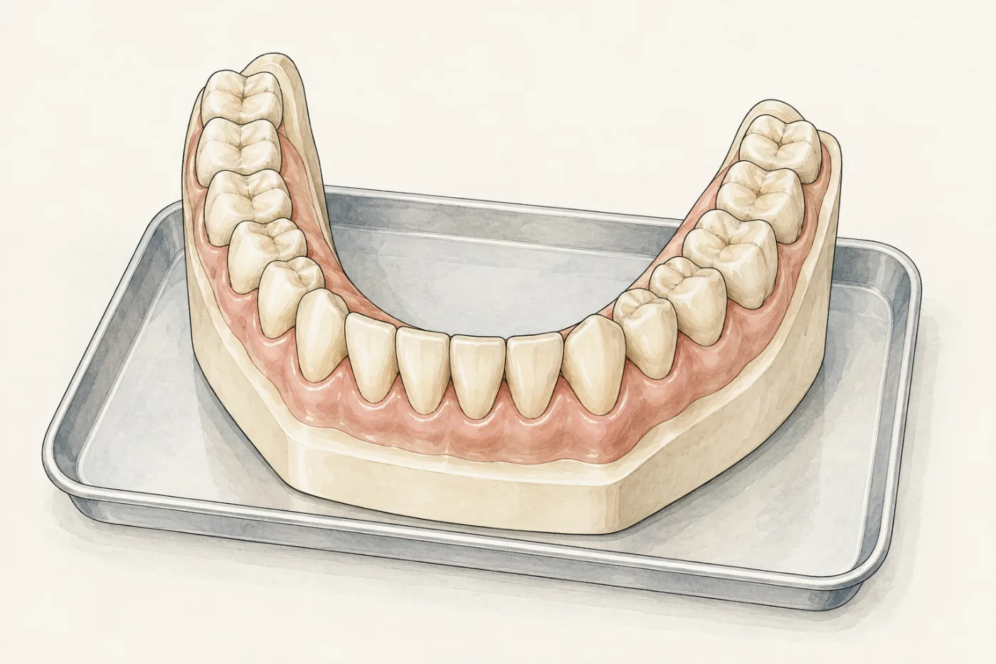
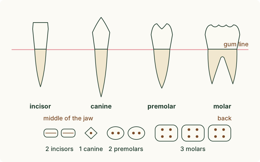
An adult has up to 32 teeth, 16 in the upper jaw and 16 in the lower jaw. Each half of each jaw, a quarter of the mouth, has the same set, from the middle to the back:
- 2 incisors: flat, chisel-shaped teeth with a sharp, straight edge, at the front;
- 1 canine: a pointed tooth, next to the incisors;
- 2 premolars: broader teeth with two bumps, called cusps, on top;
- 3 molars: the largest teeth, at the back, with a broad top and four or five cusps. The last molar, the wisdom tooth, often grows only after the age of about 17, and sometimes never.
 (enlarge)
Trap: "Canines are the big teeth at the back." Canines are pointed and stand near the front, one on each side of the incisors, four in all. The big teeth at the back are the molars.
Worked example
Naming teeth on a model
A model of a lower jaw shows, on its left side, from the middle to the back: two thin teeth with sharp, straight edges; one pointed tooth; two broader teeth, each with two cusps; three broad teeth with four or five cusps each. Name each type, and say how many teeth of each type the whole lower jaw has.
Use the shape and the place. The thin teeth with straight edges at the front are incisors. The single pointed tooth next to them is a canine. The two broader teeth with two cusps are premolars, and the three broad teeth at the back with four or five cusps are molars. The right side of the jaw is a mirror image of the left, so the lower jaw has 4 incisors, 2 canines, 4 premolars and 6 molars: 16 teeth.
Inside a tooth
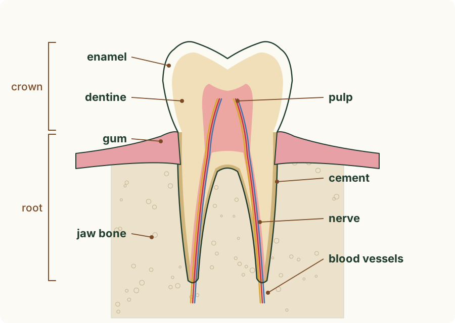
The part of a tooth above the gum is the crown; the part hidden in the jaw is the root. A tooth is built in layers:
- Enamel covers the crown. It is the hardest substance in the body, so it stands up to biting and grinding. It is not living, so a chip in the enamel cannot grow back.
- Dentine lies under the enamel and makes up most of the tooth. It is hard, like bone, but softer than enamel.
- The pulp in the middle is soft. It contains nerves, which sense pain, heat and cold, and blood vessels, which bring oxygen and nutrients to the living cells in the tooth. They enter the pulp through a small hole at the tip of each root.
- Cement covers the root and fixes the tooth firmly in the jaw.
Teeth are embedded in the jaw bone, and the gums cover the bone around the neck of each tooth.
 (enlarge)
Trap: "A tooth is dead, like a fingernail." The enamel is not living, but the pulp has living cells, nerves and blood vessels. That is why a deep crack in a tooth hurts and bleeds.
Worked example
Two broken teeth
Two players are hit in the mouth during a hockey match. Player A chips a tiny corner off an incisor and feels nothing. Player B breaks an incisor so deeply that a pink spot shows in the middle of the broken surface; it bleeds and hurts badly. Explain the difference, using the structure of a tooth.
Player A lost only a little of the outer enamel. Enamel has no nerves and no blood vessels, so there is no pain and no bleeding; but enamel is not living, so the chip will not grow back. Player B's break went through the enamel and the dentine into the pulp: the pink spot is the pulp. It contains blood vessels, so it bleeds, and nerves, so it hurts. The broken tooth stays in its place, because its root is fixed by cement in the jaw bone, under the gum.
The job of each type
Each type of tooth has a shape that suits its job in physical digestion:
- Incisors cut and bite off pieces of food, like the scalpel cutting the egg white.
- Canines grip and tear food, such as meat. In humans they are small and work with the incisors.
- Premolars and molars crush and grind food into small pieces, like the glass rod mashing the egg white. Their broad tops with cusps fit against the teeth of the other jaw.
Chewing breaks food into smaller pieces, which increases its surface area for the enzymes of chemical digestion. It also mixes the food with saliva, which makes it soft and easy to swallow.
Trap: "Molars cut food." Molars have broad, bumpy tops, not sharp edges: they crush and grind. The incisors do the cutting.
Key idea: Incisors cut and bite, canines grip and tear, premolars and molars crush and grind. A tooth has enamel on the outside, dentine under it and a pulp with nerves and blood vessels in the middle; cement fixes it into the jaw bone, under the gum.
Worked example
Eating a carrot
Describe how the teeth deal with a raw carrot, from the first bite to swallowing, and explain how this helps digestion.
The incisors cut through the carrot and bite off a piece. The tongue moves the piece to the back of the mouth, where the premolars and molars crush and grind it into small pieces, which mix with saliva. (The canines grip tough food, such as meat; a carrot hardly needs them.) This is physical digestion: the carrot is still carrot, only in smaller pieces, with no chemical change. The small pieces have a much larger surface area, so the enzymes in the gut can act on more of the food at once and digest it faster.