This part is Core content (syllabus 9.1.1, 9.2.1 and 9.2.2).
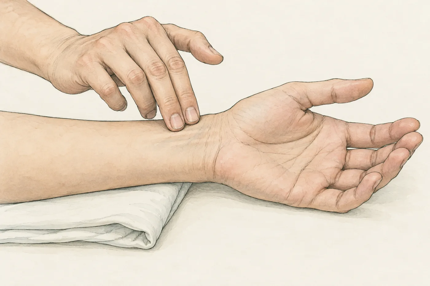
Press two fingertips gently on the inside of your wrist, just below the thumb. After a moment you feel a small push, again and again, about 75 times a minute while you sit still. Each push is a surge of blood, driven out by one beat of your heart, far away in your chest. In this module you find out how the heart drives your blood, and why each drop of it passes through the heart twice on one trip round your body.
 (enlarge)
A system of tubes with a pump
Every cell of your body needs oxygen and glucose for respiration, and makes carbon dioxide that must be taken away. Blood carries them. The circulatory system moves the blood round the body: it is a system of blood vessels (tubes) with a pump, the heart, and valves that make sure the blood flows one way only.
A valve is a set of flaps that blood can push open in one direction. If the blood starts to flow back, it pushes the flaps together and the valve shuts. The heart has four sets of valves.
Worked example
A pump without valves
The heart's muscle squeezes the blood inside it. Explain why the heart needs valves to move blood round the body.
When the muscle contracts, it squeezes the blood, and the blood is pushed out every way it can go: forwards into the arteries, but also backwards into the veins it came from. The valves shut when blood starts to flow back, so the squeezed blood can only leave forwards. With valves, each beat moves the blood one way round the body; without them, much of it would slosh back and forth.
 (enlarge)
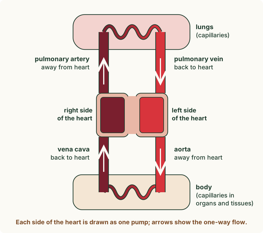
Blood that has picked up oxygen in the lungs is oxygenated: it is bright red. Blood that has given up much of its oxygen to the body's cells is deoxygenated: it is a darker red. Drawings often colour deoxygenated blood blue, but no blood is blue; in this module it is drawn dark red.
Away in arteries, back in veins
The heart pumps blood away from the heart in arteries. Blood returns to the heart in veins. Between the two, in every tissue, the blood flows through tiny vessels called capillaries, where substances pass between the blood and the cells.
Look at the diagram again ("pulmonary" means "of the lungs"). The aorta and the pulmonary artery both carry blood away from the heart, so both are arteries. The vena cava and the pulmonary vein both bring blood back, so both are veins.
Trap: "Arteries carry oxygenated blood and veins carry deoxygenated blood." The name of a vessel depends on which way the blood flows, not on its oxygen. The pulmonary artery carries deoxygenated blood away from the heart to the lungs; the pulmonary vein brings oxygenated blood back from the lungs to the heart.
Worked example
Artery or vein?
A vessel carries blood from the heart to the kidneys. Another carries blood from the lungs to the heart. Name each kind of vessel and give the reason.
The vessel to the kidneys carries blood away from the heart, so it is an artery. The vessel from the lungs brings blood back to the heart, so it is a vein, even though its blood is full of oxygen. Only the direction decides.
Inside the heart
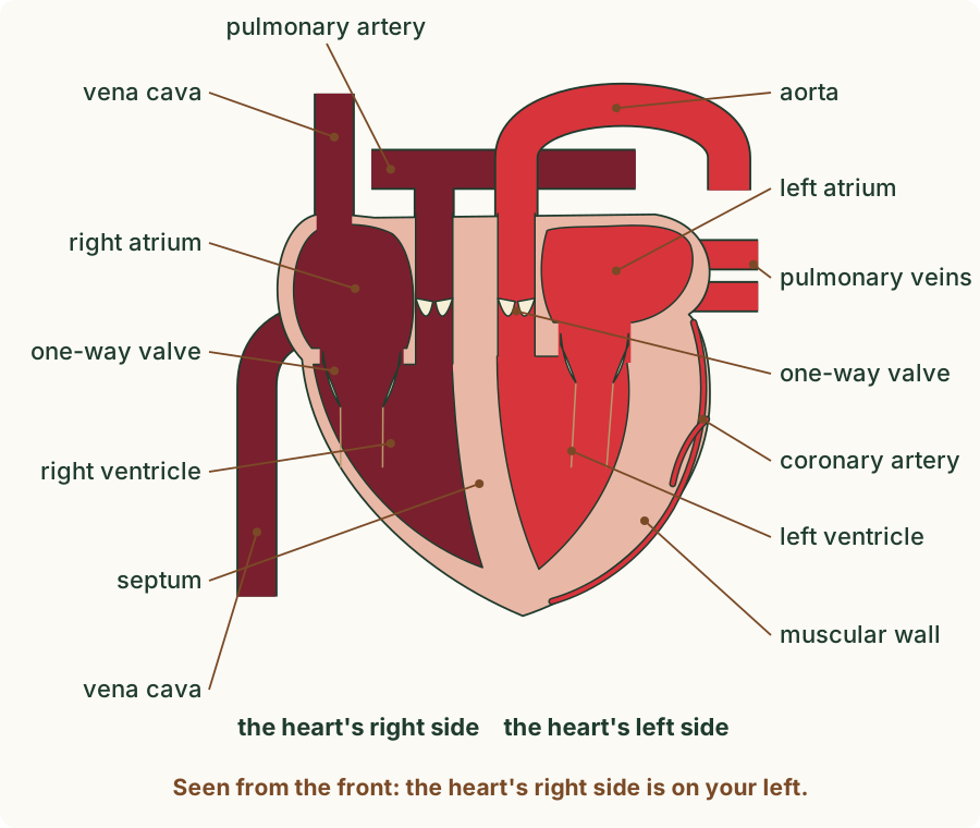
The heart is a bag of muscle about the size of your fist. Its muscular wall contracts about 75 times a minute at rest, for your whole life, without tiring. It is divided into four spaces, called chambers:
- two atria (one atrium) at the top, which receive blood coming back in the veins;
- two ventricles below them, which pump blood out into the arteries.
A wall of muscle down the middle, the septum, divides the heart into a right side and a left side. The right atrium and right ventricle hold deoxygenated blood from the body; the left atrium and left ventricle hold oxygenated blood from the lungs. One-way valves lie between each atrium and its ventricle, and where each artery leaves a ventricle.
 (enlarge)
Diagrams of the heart are drawn as if you were facing the person. So the heart's right side is on your left, and its left side on your right. The labels always name the heart's own right and left.
Trap: "The left ventricle is the one on the left of the drawing." In a diagram seen from the front, the left ventricle is on the right of the page. Find it another way: it is the ventricle with the thickest wall, and the aorta leaves it.
Worked example
Left or right?
In a diagram of a heart, the large artery that arches over the top leaves the ventricle on the right of the page, and that ventricle has the thicker wall. Which ventricle is it?
The artery that arches over the top is the aorta, and the aorta leaves the left ventricle, which has the thickest wall. The diagram is drawn from the front, so the heart's left side is on the right of the page. It is the left ventricle.
The heart's own blood supply
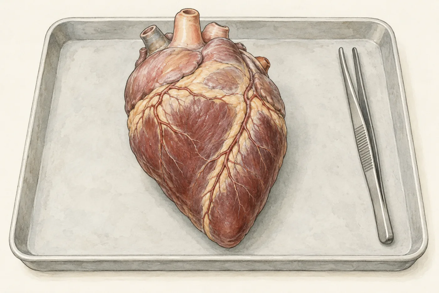
The outside of a real heart is covered by branching vessels: these are the coronary arteries. They are the first branches of the aorta, and they carry oxygenated blood into the heart muscle itself.
 (enlarge)
Worked example
Why the heart needs coronary arteries
The heart is always full of blood. Explain why its muscle still needs the coronary arteries.
The heart muscle contracts all the time, so its cells respire all the time and need a constant supply of oxygen and glucose. The blood inside the chambers is pumped straight through; it cannot reach the cells deep in the thick muscular wall. The coronary arteries branch over the surface and into the wall and bring oxygenated blood to the heart muscle cells themselves.
Key idea: The circulatory system is a system of blood vessels with a pump, the heart, and valves that keep blood flowing one way. Arteries carry blood away from the heart; veins bring it back. The heart has a muscular wall, two atria above two ventricles, a septum between its right and left sides, one-way valves, and coronary arteries that supply its own muscle.