This part is Supplement content (syllabus 9.2.7, 9.2.8, 9.2.9 and 9.2.10).
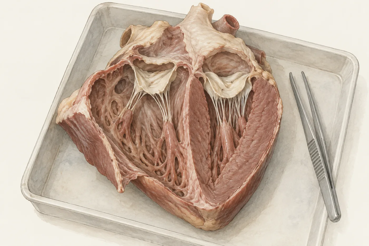
A heart from a butcher's shop, cut open from top to bottom, shows more than a diagram. The walls of its chambers are not all the same: one ventricle has a wall several times thicker than the other. Pale flaps hang at the top of each ventricle, tied down by thin white cords. In this part you find out what the walls and the flaps do in one heartbeat.
 (enlarge)
Two kinds of valve
The heart has two kinds of one-way valve.
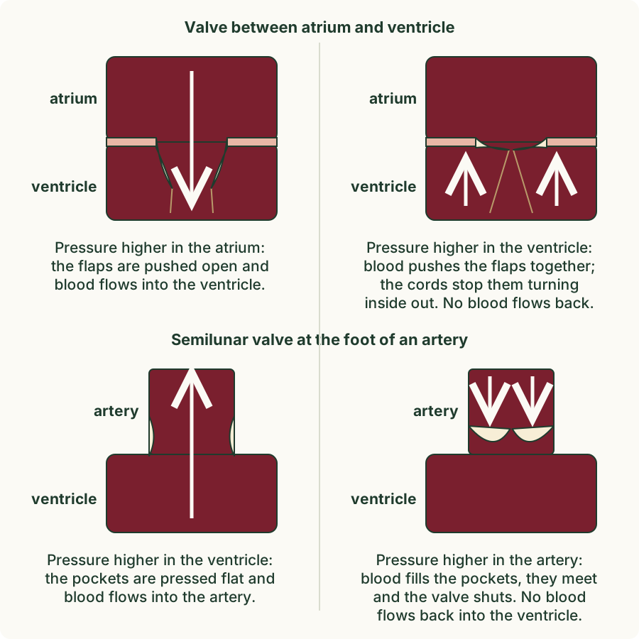
- An atrioventricular valve lies between each atrium and the ventricle below it. Its flaps open downwards into the ventricle. Thin cords tie the flaps to the ventricle wall, so they cannot turn inside out.
- A semilunar valve lies at the foot of each artery that leaves a ventricle: the pulmonary artery and the aorta. Each has three pockets shaped like half-moons, which is what "semilunar" means.
A valve has no muscle of its own. It is opened and shut by the blood around it. Blood flows from a place of higher pressure to a place of lower pressure. When the pressure is higher on the side the blood comes from, the blood pushes the valve open and flows through. When the pressure is higher on the side the blood is going to, the blood pushes the flaps together, or fills the pockets, and the valve shuts.
 (enlarge)
Trap: "The valves contract to open and close." Valves are not muscles and cannot contract. The heart's muscle changes the pressure; the pressure difference across a valve opens or shuts it.
Worked example
Which valve shuts?
The ventricles start to contract. The pressure in each ventricle rises above the pressure in the atrium above it. Which valves shut, and why does this matter?
The pressure below the atrioventricular valves is now higher than the pressure above them, so blood pushes their flaps together and they shut. The cords stop the flaps turning inside out. This stops blood being pushed back up into the atria, so when the ventricles contract the blood can only go out into the arteries.
Predict, then pump
The muscle of the atria and the muscle of the ventricles do not all contract at once. One heartbeat has four stages, and in each one some valves are open and others shut.
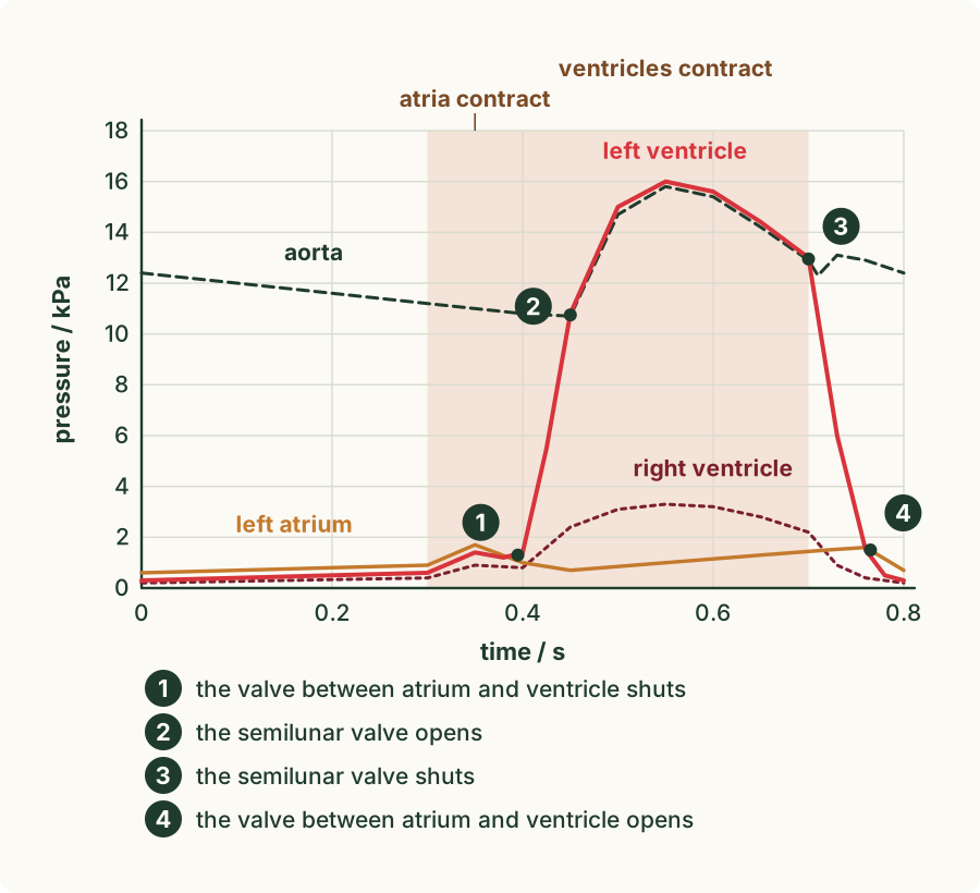
Predict first: when the heart beats, which chambers contract together? The two atria together, then the two ventricles together; the right side, then the left side; or all four at once? Make your prediction, then run the experiment below before you read on. Its graph shows pressures in kilopascals (kPa): 1 kPa is about the pressure under 10 cm of water. The pressures in this module are typical values for an adult at rest, not measurements from one person. In the model you start each contraction by tapping the chambers; in your body nobody decides: the heart starts every beat on its own.
What the model showed: four stages
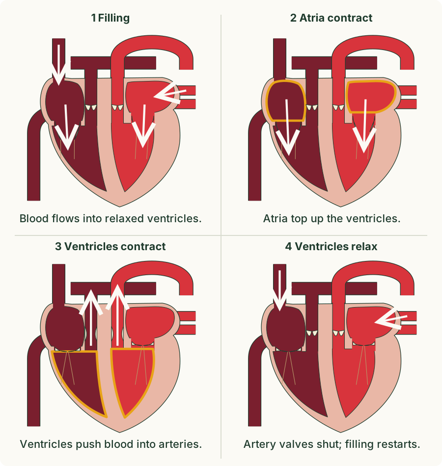
One beat of a resting heart takes about 0.8 seconds, so the heart beats about 75 times a minute. The two sides of the heart work together: both atria contract at the same time, then both ventricles.
 (enlarge)
- Filling. All the heart's muscle is relaxed. Blood flows in from the veins into both atria, and on through the open atrioventricular valves into the ventricles. The semilunar valves are shut, because the pressure in the arteries is higher than in the relaxed ventricles.
- The atria contract. The muscle of the atrial walls contracts and squeezes the last of the blood down into the ventricles.
- The ventricles contract. The pressure in the ventricles rises above the pressure in the atria, so the atrioventricular valves shut. When it rises above the pressure in the arteries, the semilunar valves open and blood is pushed out into the pulmonary artery and the aorta.
- The ventricles relax. Their pressure falls below the pressure in the arteries, so the semilunar valves shut and blood cannot flow back. When it falls below the pressure in the atria, the atrioventricular valves open and filling starts again.
 (enlarge)
Each valve opens or shuts at the moment the pressures on its two sides cross. Between points 1 and 2 on the graph both valves of the left ventricle are shut: the ventricle is squeezing, but no blood has left it yet.
Worked example
Reading the pressure graph
Use the graph. Between 0.45 s and 0.70 s, is the semilunar valve of the left ventricle open or shut? Explain using the pressures.
Between 0.45 s and 0.70 s the pressure in the left ventricle (up to 16 kPa) is higher than the pressure in the aorta, so blood pushes the semilunar valve open and flows out into the aorta. At 0.70 s the ventricle's pressure falls below the aorta's, and the valve shuts.
Thick walls and thin walls
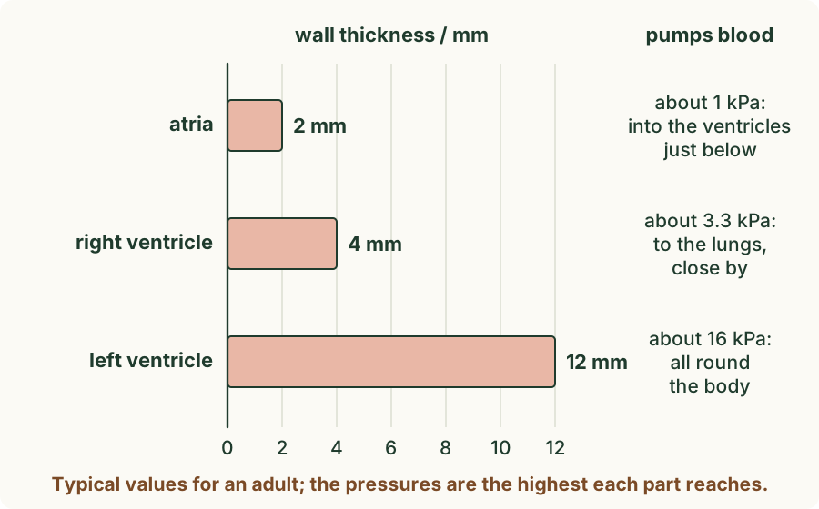
The model showed how hard each part of the heart has to push, and the walls match the work.
 (enlarge)
- The walls of the atria are thinner than the walls of the ventricles. The atria only push blood a few centimetres down into the ventricles, which are already filling.
- The wall of the left ventricle is thicker than the wall of the right ventricle. The left ventricle pumps blood all round the body, through many long vessels, so it needs a high pressure. The right ventricle pumps blood only to the lungs, close by; a lower pressure is enough, and a high pressure would damage the thin capillaries of the lungs.
More muscle can contract with more force, and makes a higher pressure.
Trap: "The left ventricle's wall is thicker because it holds more blood." Both ventricles hold and pump the same volume of blood in each beat. The left wall is thicker because it pumps the blood at a higher pressure, all round the body.
Worked example
Two ventricles
Explain why the wall of the left ventricle is thicker than the wall of the right ventricle.
The left ventricle pumps blood to the whole body, which is far away and has many vessels, so it must make a high pressure (about 16 kPa). The right ventricle pumps blood only to the lungs, close by, at a lower pressure (about 3.3 kPa). A thicker wall has more muscle, which contracts with more force.
The septum
The septum is the wall of muscle between the right and left sides of the heart. The right side holds deoxygenated blood from the body; the left side holds oxygenated blood from the lungs. The septum keeps them apart, so the blood pumped to the body is fully oxygenated and carries as much oxygen as possible to the cells.
Worked example
A hole in the septum
Some babies are born with a small hole in the septum between the ventricles. Suggest how this affects the blood that the left ventricle pumps to the body.
When the ventricles contract, some blood passes through the hole from one side to the other, so oxygenated and deoxygenated blood can mix. If deoxygenated blood reaches the left side, the blood leaving the left ventricle for the body carries less oxygen than it should, so the body's cells get less oxygen for respiration.
Key idea: (Supplement) Atrioventricular valves lie between the atria and the ventricles; semilunar valves lie at the foot of the pulmonary artery and the aorta. In one beat the atria contract together, then the ventricles contract together, then the heart relaxes and fills. Valves have no muscle: each one opens or shuts when the pressure on its two sides changes, so blood flows one way. The atria have thinner walls than the ventricles, and the left ventricle has a thicker wall than the right, because it pumps blood at a higher pressure all round the body. The septum keeps oxygenated and deoxygenated blood apart.