Cut your finger and a drop of red liquid comes out. It looks like one red liquid, but it is not. In this part you find out what blood is made of: first by spinning a tube of blood so that its parts separate, then by looking at a thin smear of blood under a microscope and counting the cells you see.
 (enlarge)
Four components
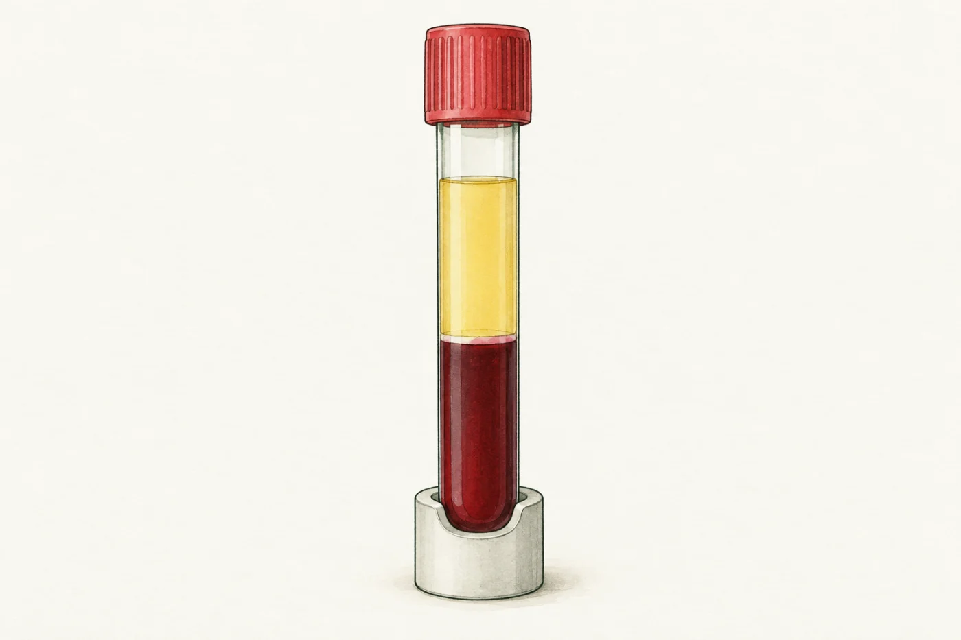
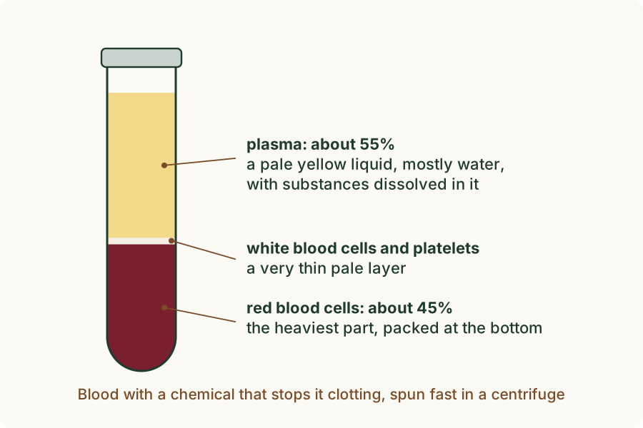
A sample of blood can be spun very fast in a centrifuge, a machine that spins tubes round so that the heavier parts sink to the bottom. A chemical is added first to stop the blood clotting (turning solid). After spinning, the tube shows layers.
 (enlarge)
 (enlarge)
- The top part, a little over half of the blood, is plasma: a pale yellow liquid, mostly water, with many substances dissolved in it.
- The bottom part, a little under half, is red blood cells, packed together. They are the heaviest part, so they sink. Their red colour comes from haemoglobin, a red protein inside them.
- Between them is a very thin pale layer. It holds the white blood cells and the platelets, small fragments of cells.
So blood has four components: red blood cells, white blood cells, platelets and plasma. The cells and platelets are carried along in the plasma.
Trap: "Plasma is water." Plasma is mostly water, but it carries many dissolved substances, such as glucose, salts, urea and hormones, as well as the blood cells.
A blood smear
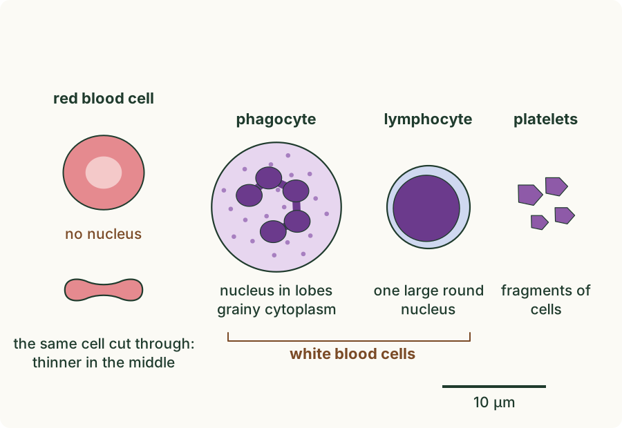
To see the cells, a drop of blood is spread very thinly across a glass slide, so that the cells lie side by side in one layer: this is a blood smear. It is dried and stained with a dye that colours a cell's nucleus purple.
 (enlarge)
- Red blood cells are by far the commonest cells. Each is a small disc, about 7.5 µm across (µm, the micrometre, is a thousandth of a millimetre). A red cell has no nucleus, and it is thinner in the middle than at the edge, so its centre looks paler.
- White blood cells are larger and much rarer. Each has a nucleus, which the stain colours purple.
- Platelets are tiny purple fragments, 2 to 3 µm across. They are pieces of cells, not whole cells.
(Supplement) There are two main kinds of white blood cell. A phagocyte is the larger kind, about 12 µm across. Its nucleus is in several lobes (rounded parts) joined together, and its cytoplasm looks grainy. The commonest phagocyte in blood is called a neutrophil. A lymphocyte is smaller, about the size of a red cell or a little bigger, with one large round nucleus that fills most of the cell and only a thin rim of cytoplasm around it. The shape of the nucleus is the best clue: a nucleus in lobes means a phagocyte, a large round nucleus means a lymphocyte.
Counting the cells
White blood cells are rarer than red ones, but how much rarer? You can find out from a smear by counting.
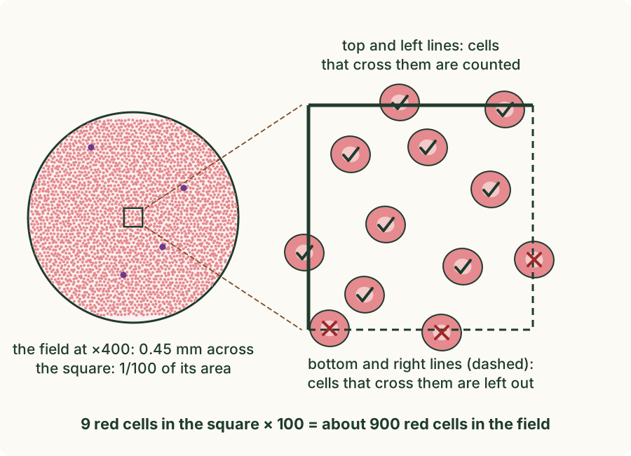
At ×400 (the ×10 eyepiece and the ×40 objective), the field of view, the circle of the slide you see, is 0.45 mm across. That is 450 µm, enough to hold about two thousand red cells. White cells are few, so you can find and count all of them in the whole field. Red cells are far too many to count one by one, so you count them in a small square instead. The square marked in the eyepiece is 40 µm along each side. Its area is one hundredth of the field's area, so the number of red cells in the whole field is about 100 times the number in the square.
A cell that lies across a side of the square needs a rule, or two people would count the same square differently. Cells that cross the top or left side are counted; cells that cross the bottom or right side are left out. Over many squares, this counts every cell on a line once and only once.
Run the experiment below before you read on. First predict how many red cells there are for each white cell; then find the white cells and count the red cells in the square.
What the counts showed
 (enlarge)
In the first field you found 4 white cells and counted 18 red cells in the square, so the whole field held about 18 × 100 = 1800 red cells. That is about 1800 ÷ 4 = 450 red cells for each white cell. The second field, a little further along the smear, gave 3 white cells and 23 red cells in its square: about 2300 ÷ 3, close to 770 red cells for each white cell.
The two fields do not agree exactly, and they should not: cells are scattered at random, so a field may happen to hold one white cell more or less. To use both fields, add the counts before you divide: (1800 + 2300) red cells ÷ (4 + 3) white cells is about 590 red cells for each white cell. All three answers, 450, 770 and 590, are the same kind of number: hundreds of red cells for each white cell. Counts from many more fields, and from many people, give a figure of several hundred red cells for each white cell in healthy blood.
A field with no white cell in it gives no ratio at all, because you cannot divide by zero. That is one reason to count more than one field.
Trap: "The smear shows how many cells are in a drop of blood." A smear shows how many cells there are of each kind compared with each other. It does not show how much blood was spread on that part of the slide, so it cannot give the number of cells in a drop.
Worked example
A spun tube
A doctor spins a tube of blood, with a chemical that stops it clotting, in a centrifuge. The tube is 80 mm tall from the bottom of the blood to its surface. The red layer at the bottom is 36 mm tall, and the pale band above it is less than 1 mm. Name each layer and work out the percentage of the blood that is red cells.
From the top: the pale yellow liquid is plasma; the thin pale band holds the white blood cells and platelets; the dark red layer at the bottom is the red blood cells, which sink because they are the heaviest part. Red cells: 36 ÷ 80 × 100 = 45% of the blood. Plasma makes up nearly all of the rest, about 55%.
Worked example
Cells in a photomicrograph
A photomicrograph is a photograph taken through a microscope. One shows many small pink discs with paler centres and no nucleus, one larger cell with a purple nucleus, and a few tiny purple specks. Identify each.
The pink discs with no nucleus are red blood cells: they are the commonest cells, and their pale centres show they are thinner in the middle. The larger cell with a purple nucleus is a white blood cell: white cells have a nucleus and are larger and much rarer. The tiny purple specks are platelets, fragments of cells.
Worked example
Red cells for each white cell
In one field at ×400 a pupil counts 3 white cells, and 21 red cells in a square that covers one hundredth of the field's area. About how many red cells are there for each white cell?
Red cells in the whole field ≈ 21 × 100 = 2100. Red cells for each white cell ≈ 2100 ÷ 3 = 700. The answer has no unit: it is a number of cells for each cell. One field gives only a rough answer; counting several fields and adding the counts gives a better one.
Worked example
Lymphocyte or phagocyte?
(Supplement) Two white cells are seen in a smear. Cell A is about 12 µm across, with a nucleus in four lobes and grainy cytoplasm. Cell B is about 8 µm across, with one large round nucleus and a thin rim of clear cytoplasm. Identify each cell and give the evidence.
Cell A is a phagocyte: its nucleus is in lobes and its cytoplasm is grainy, and it is the larger of the two. Cell B is a lymphocyte: its nucleus is large and round and fills most of the cell. The shape of the nucleus is the main evidence; size alone could mislead, because a lymphocyte can sometimes be fairly large.
Key idea: Blood has four components: red blood cells, white blood cells, platelets and plasma. In a stained smear, red cells are small pink discs with no nucleus; white cells are larger, much rarer, and have a nucleus; platelets are tiny fragments. There are hundreds of red cells for each white cell. (Supplement) A phagocyte has a nucleus in lobes and grainy cytoplasm; a lymphocyte has one large round nucleus that fills most of the cell.