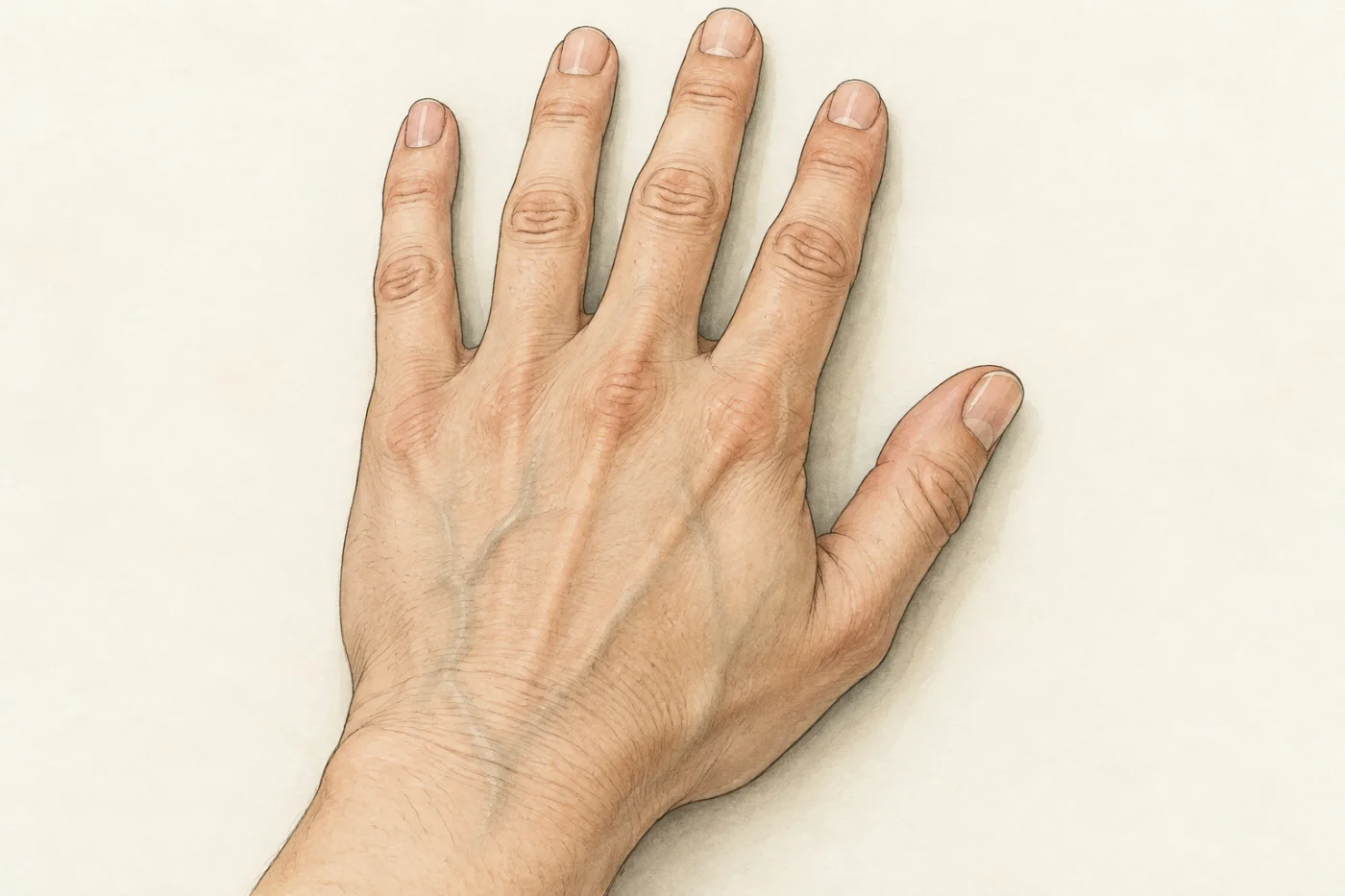
Press two fingers lightly on the inside of your wrist and you feel a pulse: blood is pushed along a vessel in surges (strong pushes), one for each beat of your heart. Now look at the back of your hand. The vessels you can see under the skin carry blood too, but you feel no pulse in them. In this part you find out what makes these two kinds of vessel different, by looking at both under a microscope and measuring them.
 (enlarge)
Three kinds of blood vessel
Blood leaves the heart in arteries: vessels that carry blood away from the heart. Arteries branch again and again into smaller and smaller vessels, and the smallest of all are the capillaries, which run between the cells of every tissue. Capillaries join up again into larger vessels, the veins, which carry blood back to the heart.
So the names say which way the blood goes, not what is in it. The pulse in your wrist is in an artery, the radial artery. The vessels on the back of your hand are veins.
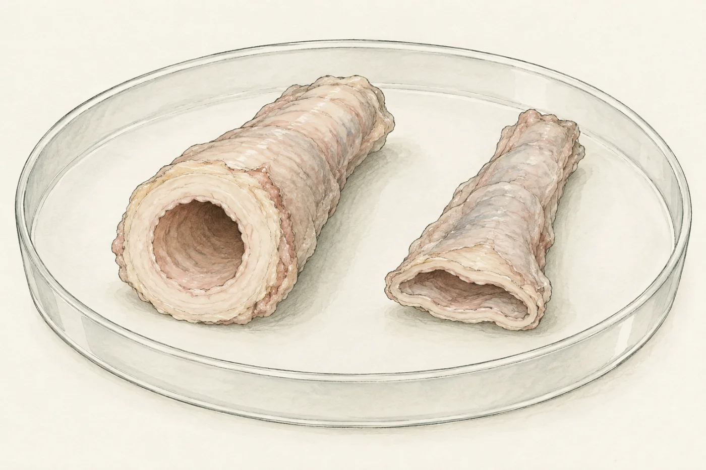
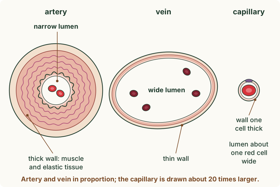
Every blood vessel is a tube. The space inside the tube, where the blood flows, is called the lumen. Around the lumen is the wall of the vessel. An artery's wall and a vein's wall both contain muscle and elastic tissue (tissue with fibres that stretch and then spring back), lined on the inside by a layer of cells one cell thick. What differs is how much of each there is.
 (enlarge)
Cut out of the body, an artery keeps its round shape: its thick wall holds the lumen open. A vein of a similar size has a thin wall, and it flattens. But a vessel can be squashed while a slide is made, so shape alone does not tell you which is which. Under the microscope you look at the wall: how thick it is compared with the lumen, and whether it has a thick layer of muscle and wavy elastic fibres.
Free play: nothing here is marked or saved
Pulse in the wrist, none in the hand
Each beat of the heart pushes a surge of blood into the arteries. Where can two fingers feel the surges? The clipboard draws the blood’s pressure in kilopascals (kPa).Supplement
Still picture: your device asks for less motion.
Try: Speed the heart up to 140 a minute, as after a run. What do the two fingers feel now?
- Artery, wrist (kPa)
- 10.5–15.5
- Capillaries, fingertip (kPa)
- 3
- Vein, back of hand (kPa)
- 1
- Pulse under the fingers
- 70 a minute
- The pulse is the surge each heartbeat sends along the arteries. Count the pulses at the wrist, on the palm side below the thumb, for a minute and you have the heart rate.
- The pressure in an artery rises with each surge and falls between them. By the capillaries the surges have died away: the pressure there is lower and steady.
- In the veins the pressure is lowest and steady, so there is no pulse to feel on the back of the hand.
About this model. A model with illustrative pressures for a resting adult, the reading’s typical values: here they stay the same at every heart rate, though in life the pressure in the arteries rises in exercise. The surges are slowed down a great deal so you can follow them; in life one reaches the wrist in less than a tenth of a second. The vessels are drawn as plain lines, fewer and simpler than they are, and their walls are not drawn. Veins are not blue: the colours only tell the vessels apart. You would feel a wrist pulse on the palm side, below the thumb; this hand lies palm down so the veins on its back show.
Measuring a vessel on a slide
A transverse section is a thin slice cut straight across a vessel, so that you see it as a ring. You will look at a stained slide with an artery and a vein side by side in it.
The microscope's field of view is the circle of the specimen you see. With the ×10 eyepiece and the ×10 objective (×100 in all), the field of view on this microscope is 1.8 mm across, which is 1800 µm (1 mm = 1000 µm; µm is the micrometre, a thousandth of a millimetre). Because you know how wide the whole field is, you can measure anything in it as a fraction of the field.
To compare two vessels of different sizes, you do not compare their walls alone: a big vessel may have a thicker wall just because it is big. Instead you compare the wall with the lumen. Divide the wall thickness by the lumen's width:
wall ÷ lumen = wall thickness ÷ width of the lumen
Both are lengths in µm, so the units cancel and the result has no unit. A large value means a thick wall for the size of the lumen; a small value means a thin wall around a wide lumen.
Run the experiment below before you read on. First decide which vessel is the artery, from what you see in the wall; then measure both.
What the measurements showed
 (enlarge)
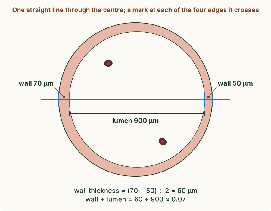
Your line crossed four edges: the outer and inner edge of the wall on one side, then the inner and outer edge on the other. Wall thickness is the mean of the two sides, because a section is rarely cut perfectly straight across. The lumen is the distance between the two inner edges.
For artery X, the first artery you measured, the wall was about as thick as the lumen was wide, so wall ÷ lumen was close to 1. For vein Y it was well under 0.1. The second pair of vessels gave the same pattern, with different numbers. Keep the arteries and veins apart when you take a mean: a mean of an artery and a vein together describes neither vessel.
Your numbers come from one slide, not from every artery and vein in the body. They also depend on how flat each section was cut. That is why you measured along one line through the centre of each ring, and why a second pair matters: if both pairs show the same difference, it is more likely to be real.
 (enlarge)
| artery | vein | capillary | |
|---|---|---|---|
| wall | thick, with a thick layer of muscle and elastic tissue | thin, with little muscle and elastic tissue | one cell thick |
| lumen | narrow for the size of the vessel | wide | very narrow, about the width of one red blood cell |
| valves | none along its length | valves along its length | none |
Trap: "Veins are blue." Blood in veins is dark red. Veins look blue-green through the skin because of the way skin scatters light, and diagrams colour them blue only to tell them apart from arteries.
Worked example
Which is the artery?
A section shows two vessels. Vessel X has a ring of wall about 250 µm thick around a lumen about 300 µm wide; its wall has a thick layer of muscle with wavy pink fibres. Vessel Y has a wall about 40 µm thick around a lumen about 900 µm wide, and its lumen is slightly flattened. Which vessel is the artery? Give two pieces of evidence.
Look at the wall, not the shape. X is the artery: its wall is thick compared with its lumen (wall about 250 µm, lumen about 300 µm), and the wall has a thick layer of muscle with wavy elastic fibres. Y has a thin wall around a wide lumen, so Y is the vein. The flattened shape of Y fits a vein, but it could also come from the way the slide was cut, so it is not good evidence on its own.
Worked example
Working out wall ÷ lumen
On a scaled photograph of a vein, a line through its centre crosses the wall on the left over 70 µm and on the right over 50 µm. The lumen between the inner edges is 900 µm. Calculate the wall thickness and wall ÷ lumen.
Wall thickness is the mean of the two sides: (70 + 50) ÷ 2 = 60 µm. Then wall ÷ lumen = 60 ÷ 900 = 0.067, about 0.07. The answer has no unit, because both lengths were in µm. A value this small fits a vein: a thin wall around a wide lumen.
Valves in veins
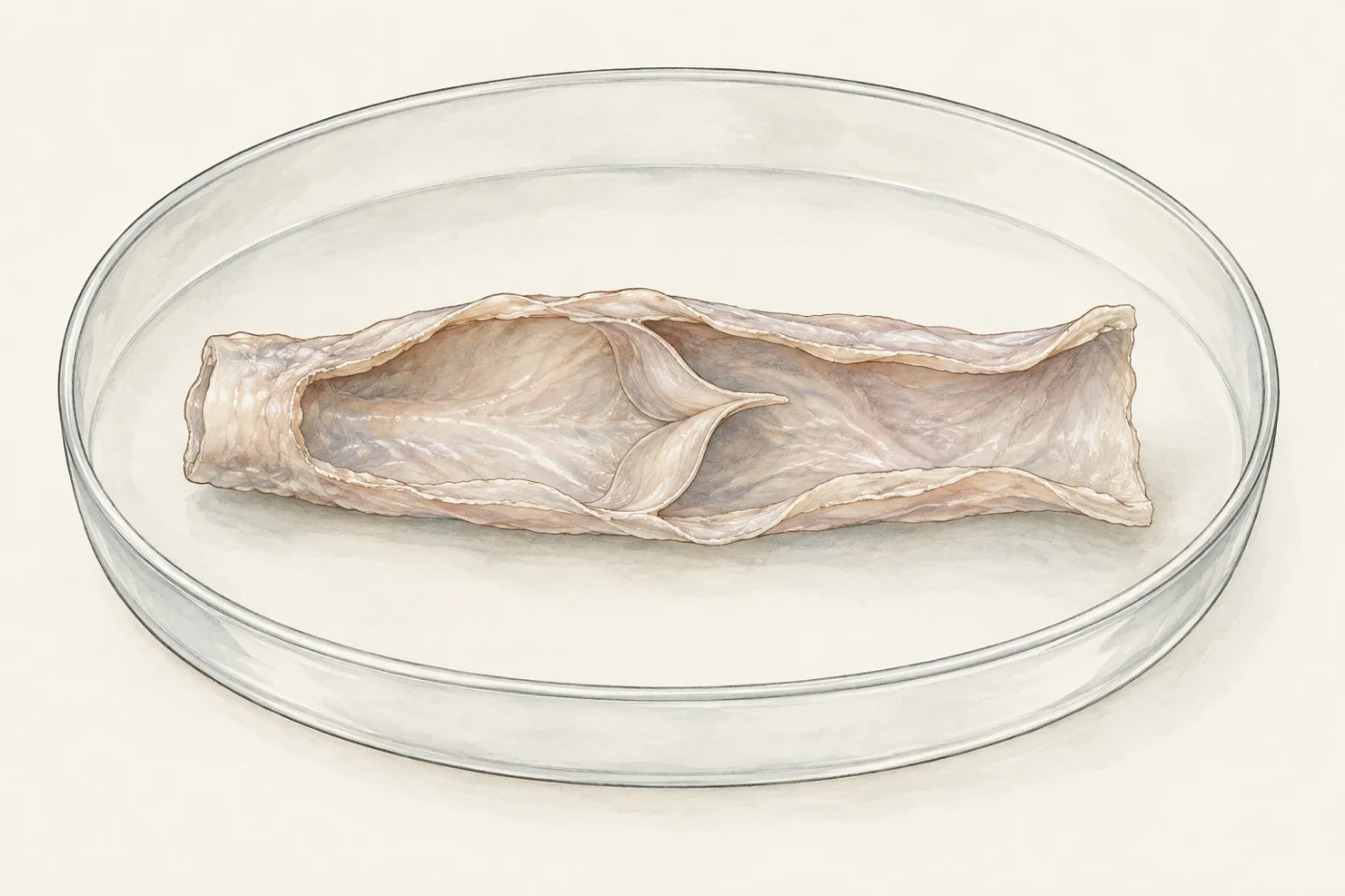
Some veins have valves: pairs of thin flaps, each shaped like a pocket, attached to the inside of the wall. The flaps point towards the heart.
 (enlarge)
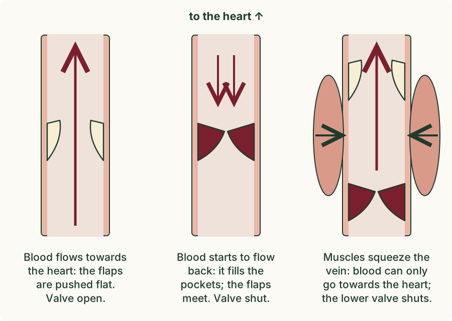
When blood flows towards the heart, it pushes the flaps flat against the wall and the valve is open. If blood starts to flow back, it fills the pockets; the flaps swing out and meet in the middle, and the valve shuts. So valves prevent the backflow of blood: blood in a vein can only go one way, towards the heart. Arteries have no valves along their length.
 (enlarge)
Many veins, such as those in the legs, run between muscles. When the muscles contract, they squeeze the veins. The valves make sure the squeezed blood goes towards the heart, not away from it. This is one reason why walking helps to return blood from the legs.
Trap: "Arteries have valves to stop the blood flowing back." The only valves on the arteries are where they leave the heart. Along their length, the pressure from the heart keeps the blood moving forwards, so arteries do not need valves.
Worked example
Why only veins need valves
(Supplement) Explain why veins have valves but arteries do not.
The blood in an artery has just been pushed out of the heart at high pressure, and the pressure keeps it moving away from the heart. By the time blood has passed through the capillaries, its pressure is very low. At such a low pressure, blood in a vein could flow backwards, for example down a leg when you stand up. The valves in veins prevent this backflow, so the blood keeps moving towards the heart.
Pressure and the structure of a wall
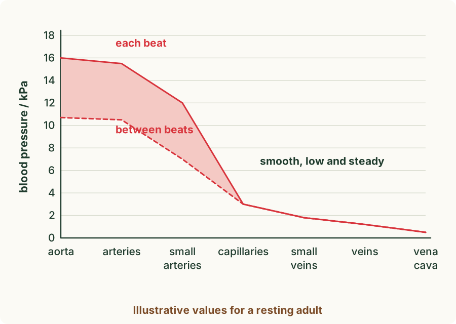
(Supplement) With each beat, the heart pushes blood into the arteries at high pressure. The pressure rises with each beat and falls between beats: this is the pulse you feel in your wrist.
 (enlarge)
The values on the graph are typical for a resting adult; the kPa (kilopascal) is a unit of pressure. Blood pressure is highest in the arteries near the heart and falls along the path. In the arteries it rises and falls with each beat; in the capillaries and veins it is low and steady.
The structure of each vessel fits the pressure of the blood it carries.
- An artery has a thick wall, which can withstand the high pressure without bursting.
- The elastic tissue in an artery's wall stretches as each surge of blood arrives and recoils (springs back) between beats. This evens out the surges, so the flow becomes smoother further from the heart.
- The muscle in an artery's wall can contract or relax, which narrows or widens the artery and changes how much blood flows to an organ.
- A vein carries blood at low pressure, so it does not need a thick wall. Its thin wall and wide lumen let blood flow back to the heart easily, with little to slow it down.
- Valves prevent the backflow of this low-pressure blood, and the contractions of the muscles around the veins help to push it towards the heart.
Trap: "Arteries always carry oxygenated blood." An artery is any vessel that carries blood away from the heart. The pulmonary artery carries deoxygenated blood from the heart to the lungs.
Worked example
Harvey's finger
In 1628 the doctor William Harvey described a simple test on the veins of the forearm. With a band round the upper arm, the veins on the forearm stood out, with small swellings along them where the valves were. Harvey pressed one finger on a vein, so that no blood could come up it from the hand. With a second finger he stroked the blood along the vein towards the heart, past the next valve. The stretch of vein between the first finger and that valve stayed empty, and filled again only when he lifted the first finger. When he tried to stroke blood the other way, away from the heart, he could not push it past a valve. What did this show, and how does it fit the structure of a vein?
Blood could be pushed through a valve towards the heart, but not back through it. The empty stretch stayed empty because the valve above it shut and stopped the blood flowing back into it; it filled only from below, from the hand. This showed that blood in the veins flows only towards the heart, and that the valves prevent backflow. It fits the structure: the flaps of each valve point towards the heart, so blood flowing that way opens them and blood flowing back shuts them.
Key idea: Arteries carry blood away from the heart and veins carry it back. An artery has a thick wall of muscle and elastic tissue and a narrow lumen; a vein has a thin wall, a wide lumen and valves that prevent backflow. (Supplement) The thick, elastic wall of an artery withstands the high pressure from the heart and smooths the flow; the blood in veins is at low pressure, so veins have thin walls and wide lumens, and need valves and the squeezing of muscles to keep the blood moving towards the heart.