Arteries and veins are big enough to see in a section at ×100. But no cell in your body takes anything straight from an artery or gives anything straight to a vein. Every exchange between the blood and a cell happens in vessels far too small to see at ×100. In this part you turn to a higher magnification to find them, and work out why they are built the way they are.
Too small for ×100
Between the arteries and the veins, the blood flows through capillaries. A capillary is about 8 µm across, about the width of one red blood cell (a red blood cell is about 7 µm across). At ×100, where the whole field is 1800 µm wide, a capillary would be less than half a percent of the field: you might see a red blood cell in it, but its wall is far too thin to see.
Turn the nosepiece to the ×40 objective: with the ×10 eyepiece, that is ×400. The field of view is now 0.45 mm (450 µm) across, a quarter of the ×100 field, so everything looks four times wider. Look in the tissue around the vessels for tiny vessels with a red blood cell inside. Then run the part of the experiment below, which starts at this point, before you read on.
What the ×400 field showed
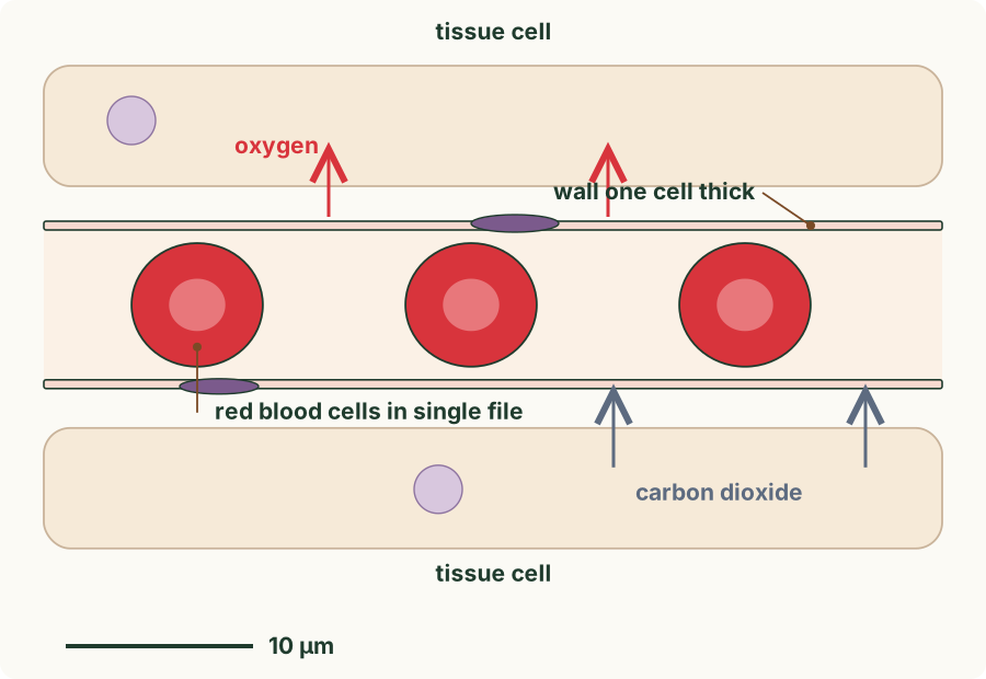
In the tissue around the large vessels there were tiny vessels, each about as wide as the red blood cell inside it. A red blood cell in a capillary almost fills the lumen, so the cells pass along in single file, one after another.
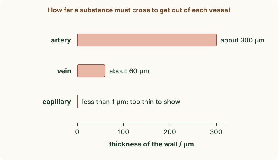
The wall of a capillary is not a ring of muscle. It is a single layer of very thin, flattened cells: the wall is one cell thick, less than 1 µm. It has no muscle and no elastic tissue, and no valves.
 (enlarge)
Trap: "Capillaries are tiny arteries." Capillaries are a different kind of vessel. An artery, however small, has muscle in its wall; a capillary's wall is a single layer of thin cells with no muscle.
Worked example
The path of the blood
A red blood cell leaves the heart and goes to a muscle in the leg, then back to the heart. Name the three kinds of vessel it passes through, in order, and say what happens in each.
It goes through an artery first: arteries carry blood away from the heart, under high pressure. The arteries branch into capillaries inside the muscle, where substances are exchanged between the blood and the muscle cells. The capillaries join into veins, which carry the blood back to the heart at low pressure.
What capillaries do
 (enlarge)
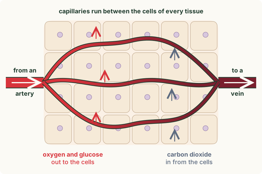
Capillaries form a network, a capillary bed, between the cells of every tissue. No cell is far from a capillary. Capillaries have two functions:
- They carry blood from the arteries to the veins, close to every cell.
- They let substances be exchanged between the blood and the cells around them.
Oxygen and glucose pass out of the blood, through the capillary wall, to the cells, which use them in respiration. Carbon dioxide, made by respiration, passes the other way, into the blood. Other substances are exchanged too: amino acids and other nutrients pass out to the cells, and waste such as urea passes into the blood.
These substances move by diffusion (the net movement of particles from where they are more concentrated to where they are less concentrated). Oxygen is more concentrated in the blood arriving from an artery than in the cells, which keep using it, so it diffuses out. Carbon dioxide is more concentrated in the cells, which keep making it, so it diffuses into the blood.
Trap: "Red blood cells pass out of the capillaries to give oxygen to the cells." The red blood cells stay inside the capillaries. Oxygen leaves the red blood cells and diffuses through the capillary wall to the cells.
Worked example
How far oxygen must go
The wall of the artery on your slide was about 300 µm thick. A capillary's wall is about 0.5 µm thick. How many times further would oxygen have to diffuse to get through the artery's wall?
300 ÷ 0.5 = 600 times further. Diffusion over a few micrometres is quick; diffusion over hundreds of micrometres is far too slow to supply cells. So substances are exchanged through the thin walls of capillaries, not through the thick walls of arteries or veins.
Structure and function
(Supplement) Each feature of a capillary fits its job of exchange.
 (enlarge)
- The wall is one cell thick, so the diffusion distance (the distance a substance must travel) between the blood and the cells is very short, and diffusion is fast.
- The lumen is narrow, about the width of one red blood cell. The red blood cells squeeze through in single file, close to the wall, so the oxygen they carry has only a short way to go. The blood also moves slowly through the huge number of capillaries, giving more time for exchange.
- Capillaries are many and branch between the cells, so their walls give a very large surface area for exchange, and every cell is close to one.
- The thin wall lets some of the plasma (the liquid part of blood) leak out between the cells of the wall, bringing dissolved substances to the cells around them.
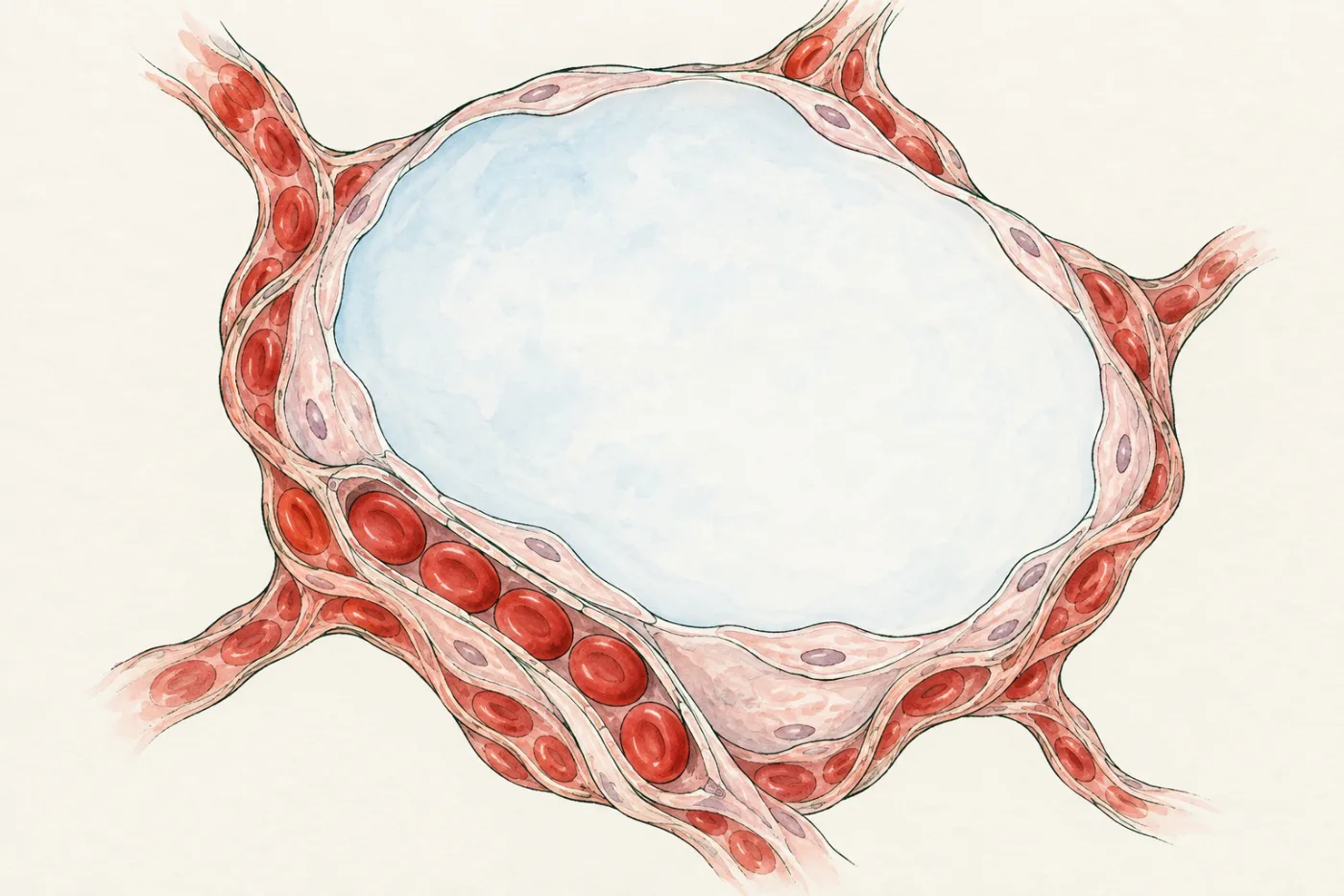
The same features are used wherever substances enter or leave the blood. In the lungs, capillaries are wrapped round the air sacs, so oxygen diffuses from the air into the blood across two walls, each one cell thick.
 (enlarge)
Worked example
A narrow lumen
(Supplement) Explain how the narrow lumen of a capillary helps oxygen to reach the cells of a tissue.
The lumen is about the width of one red blood cell, so the red blood cells pass through in single file, pressed close to the wall. Oxygen leaving a red blood cell then has only a short distance to diffuse, through the thin wall, to the cells. The blood also moves slowly through the huge number of capillaries, which gives more time for oxygen to diffuse out.
Worked example
More capillaries
(Supplement) Muscle that is trained for long-distance running has more capillaries in each square millimetre than the same muscle in a person who does not train. Suggest why this helps the runner.
More capillaries give a larger surface area for exchange, and each muscle cell is closer to a capillary. So oxygen and glucose diffuse from the blood to the muscle cells faster, and carbon dioxide is removed faster. The muscle cells can respire aerobically for longer.
Key idea: Capillaries are the smallest vessels, about the width of one red blood cell, with a wall one cell thick. They carry blood between the arteries and the veins, close to every cell, and they are where substances are exchanged: oxygen and glucose pass out to the cells, and carbon dioxide passes into the blood. (Supplement) The wall one cell thick gives a short diffusion distance; the narrow lumen keeps the red blood cells close to the wall; their great number gives a large surface area and keeps every cell close to a capillary.