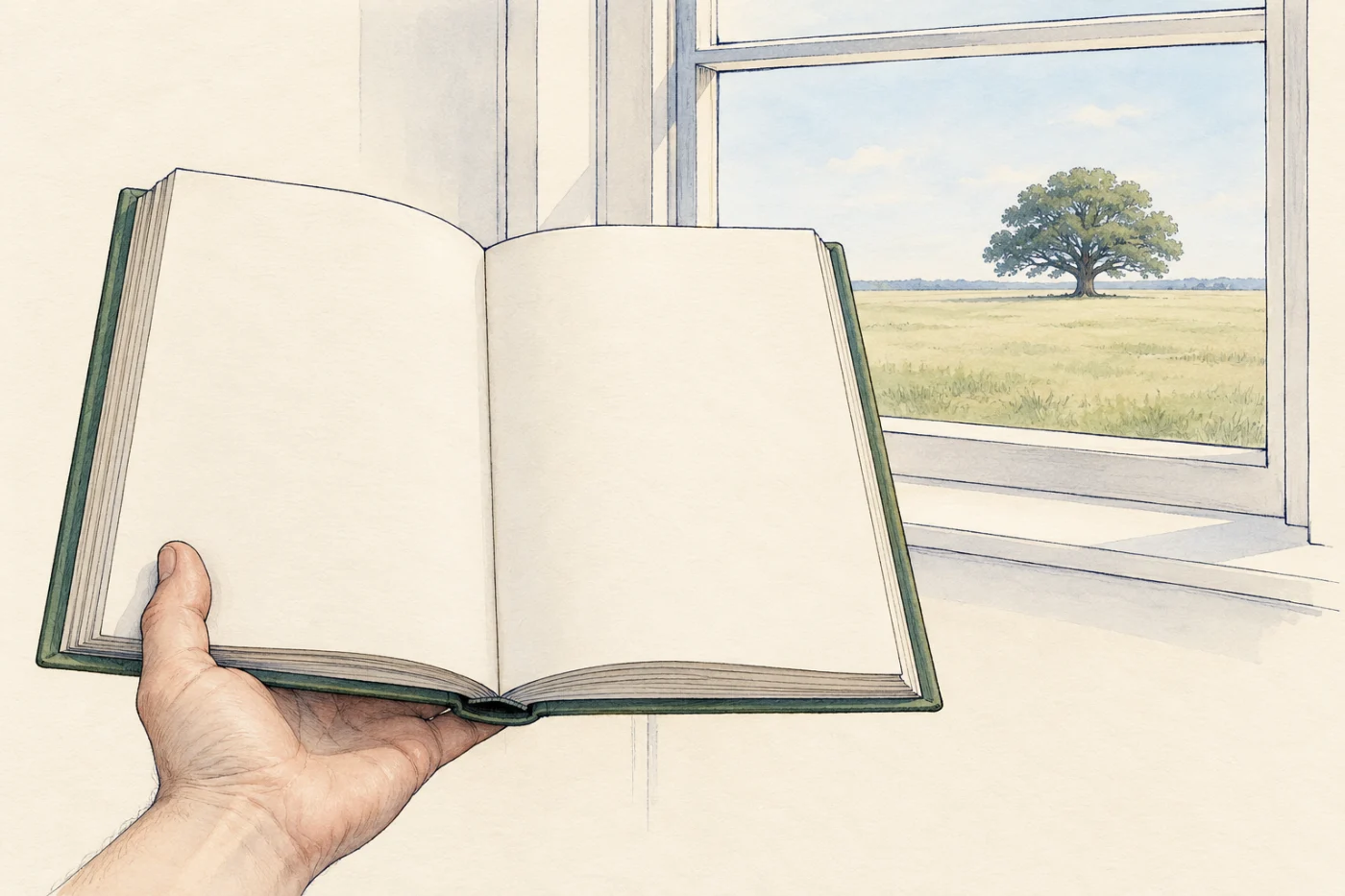
You look up from a book to a tree outside the window, and both are sharp and full of colour. A moment later someone switches on a bright lamp, and you can still see the page. Your eye does all of this without you deciding anything. In this part you meet the sense organs, find each part of the eye in a diagram, and follow the light from an object to the place where the eye detects it.
 (enlarge)
Sense organs
In the module on nerves and reflexes you met receptors: cells that detect a stimulus and start impulses in a sensory neurone. Receptor cells are often grouped together in sense organs. A sense organ is a group of receptor cells that respond to a specific stimulus: each kind of sense organ detects one kind of change.
| Sense organ | The stimulus its receptors respond to |
|---|---|
| eye | light |
| ear | sound |
| skin | touch and temperature |
| tongue | chemicals (in food and drink) |
| nose | chemicals (in the air) |
The receptor cells in the eye respond to light and do not respond to sound; the receptors in the ear respond to sound and not to light. Whichever sense organ detects a stimulus, its receptors start impulses that travel along sensory neurones to the central nervous system.
Trap: "A sense organ is one receptor." A sense organ is a group of many receptor cells, all responding to the same kind of stimulus. The retina of one eye holds millions of light receptors.
The parts of the eye
 (enlarge)
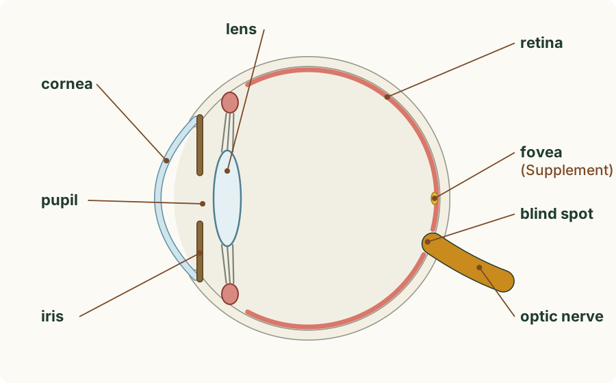
The diagram shows an eye cut through from front to back, as if seen from the side. Light comes in from the left. Starting at the front:
- The cornea is the clear, curved window at the very front of the eye.
- The iris is the coloured ring of muscle just behind the cornea. In a cut-through diagram you see it as two short halves, one above and one below.
- The pupil is the gap in the middle of the iris: a hole that light passes through. In the diagram it is the space between the two halves of the iris.
- The lens is the clear oval part just behind the pupil.
- The retina is the layer that lines the back of the eye. It contains the light receptors.
- The optic nerve is the thick bundle of neurones that leaves the back of the eye and runs to the brain.
- The blind spot is the place on the retina where the optic nerve leaves the eye.
The eye also has a tough white outer layer and is filled with clear jelly. They are drawn in the diagrams, but you do not need their names.
 (enlarge)
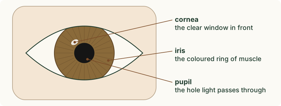
From the front you see the same parts face on: the iris as a coloured ring and the pupil as a black circle in its middle. The pupil looks black because light goes in through it and very little comes back out.
Trap: "The pupil is the coloured part of the eye." The coloured part is the iris. The pupil is the hole in the middle of the iris, which is why it looks black.
Following the light
 (enlarge)
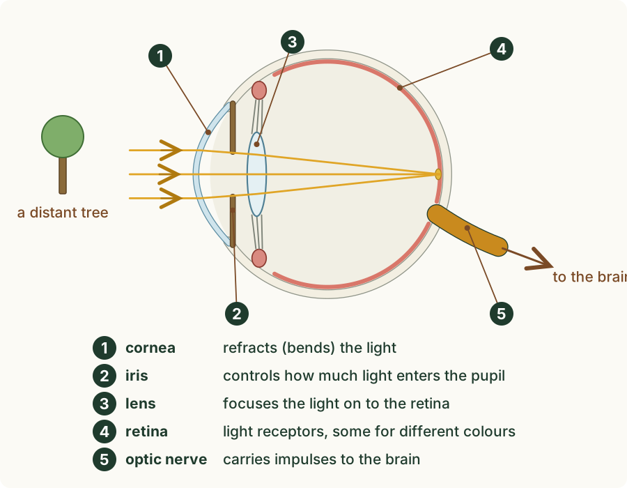
Light from an object passes through the parts of the eye in order, and each part has its own job.
- The cornea refracts light: it bends the rays as they enter the eye. ("Refract" means to bend light as it passes from one material into another.)
- The light passes through the pupil. The iris controls how much light enters the pupil, by making the pupil wider or narrower.
- The lens focuses light on to the retina: it bends the rays again so that they meet on the retina and form a sharp image there.
- The retina contains light receptors. Some of them are sensitive to light of different colours. When light falls on them, they start impulses.
- The optic nerve carries impulses to the brain.
The image is formed on the retina, not on the lens or in the brain. The brain receives impulses from the retina along the optic nerve.
Trap: "The lens does all the bending of the light." The cornea refracts the light first, as it enters the eye. The lens then bends it again to focus it on the retina.
Run the experiment below before you read on. First predict where the light from the book forms an image. Then tap each part of a large model eye from a clue about its job or its place; its name appears once you have found it. At the end there is a short, optional test you can do on your own eye. The rays in the model are drawn lines: light itself cannot be seen as it travels through the eye.
What the experiment showed
The light from the book passed through the cornea, the pupil and the lens, and met on the retina at the back of the eye: that is where the image forms. Each part you tapped has one job on that path. The cornea and the lens bend the light, the iris sets how much light gets through the pupil, the retina detects the light, and the optic nerve carries impulses away to the brain.
The blind spot
At the blind spot, the neurones from all over the retina gather and leave the eye as the optic nerve. There is no room for light receptors there, so the blind spot has no light receptors. Light that falls on it starts no impulses, and you cannot see the part of the scene whose light lands there.
You do not usually notice it, because your two eyes see the scene from slightly different places: light that falls on the blind spot of one eye falls on light receptors in the other. With one eye closed you can find it. In the experiment's optional test, the dot vanished when its light fell on your blind spot. If it never vanished for you, that is common: it depends on the screen's size and your distance from it, and it is not a sign of a problem with your eye.
Trap: "The blind spot is a damaged part of the eye." Every healthy eye has a blind spot. It is simply the place where the optic nerve leaves, with no light receptors.
Worked example
Naming a part from its job
A student is asked to name: (a) the clear window that refracts light as it enters the eye; (b) the part that controls how much light enters; (c) the layer that contains light receptors; (d) the part that carries impulses to the brain.
(a) The cornea: it is at the very front and refracts the light. (b) The iris: its muscles change the size of the pupil. (c) The retina: it lines the back of the eye. (d) The optic nerve. Check each answer against the order of the light's path: cornea, pupil, lens, retina, then the optic nerve to the brain.
Worked example
The order of the path
Put these in the order that light, and then the impulses, reach them: lens, optic nerve, cornea, retina, pupil.
Light enters through the cornea, passes through the pupil and then the lens, and falls on the retina. The retina's receptors start impulses that travel along the optic nerve. So the order is cornea → pupil → lens → retina → optic nerve. The optic nerve comes last because it carries impulses, not light.
Worked example
Which sense organ?
Name the sense organ that detects each stimulus: (a) the smell of toast; (b) a hot radiator under your hand; (c) a whistle; (d) the sourness of a lemon.
(a) The nose: its receptors respond to chemicals in the air. (b) The skin: its receptors respond to temperature. (c) The ear: it responds to sound. (d) The tongue: it responds to chemicals in food and drink. Each sense organ is a group of receptor cells that respond to one kind of stimulus.
Worked example
Light on the blind spot
Explain why you cannot see an object whose light falls on the blind spot of one eye.
The blind spot is where the optic nerve leaves the eye, and it has no light receptors. Light falling there does not start any impulses, so no impulses reach the brain from that part of the image, and the object is not seen by that eye. Light that falls on the rest of the retina, which does have receptors, is seen.
Key idea: A sense organ is a group of receptor cells that respond to one kind of stimulus: the eye light, the ear sound, the skin touch and temperature, the tongue and the nose chemicals. In the eye, the cornea refracts light; the iris controls how much light enters through the pupil, the hole in its middle; the lens focuses light on to the retina; the retina contains light receptors, some sensitive to different colours; the optic nerve carries impulses to the brain. The blind spot, where the optic nerve leaves, has no light receptors.