This part is for the Supplement (extended) paper. You can read a page held close to your face and, a moment later, see a tree far away just as sharply. The rays of light from the two arrive at your eye very differently, yet both images end up sharp on the retina. In this part you find out how the eye changes its focus, where the two kinds of light receptor sit in the retina, and why you look straight at something to see it in detail.
 (enlarge)
Focusing near and far: accommodation
 (enlarge)
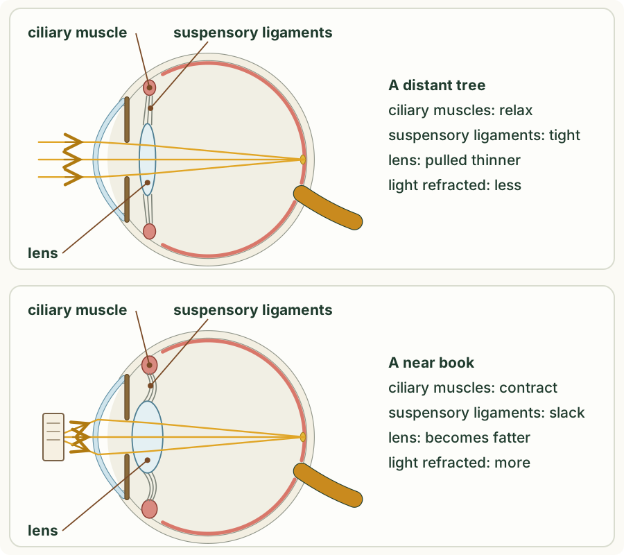
Rays of light from a distant object are almost parallel when they reach the eye, but rays from a near object are still spreading out, so they need to be bent more to meet on the retina. The cornea's shape does not change, and it refracts the light by the same amount every time, so it is the lens that changes: a fatter lens refracts light more, a thinner lens less. Changing the shape of the lens to focus on near or distant objects is called accommodation.
The lens is elastic: like a rubber ball, it springs back to its own rounder shape when nothing pulls on it. It hangs in a ring of ciliary muscle on fine threads called suspensory ligaments.
- Distant object: the ciliary muscles relax. The ring of muscle gets wider, so the suspensory ligaments are pulled tight. They pull on the edge of the lens and make it thinner. A thinner lens refracts the light less, and the nearly parallel rays meet on the retina.
- Near object: the ciliary muscles contract. The ring of muscle gets smaller, so the suspensory ligaments slacken. With no pull on its edge, the elastic lens springs into a fatter, rounder shape. A fatter lens refracts the light more, and the spreading rays meet on the retina.
In a real eye this happens automatically, every time you look from something near to something far. In the experiment's model, you change the lens yourself to see what the eye has to do.
Trap: "The ciliary muscles contract to pull the lens fat." Contracting ciliary muscles let the suspensory ligaments slacken: nothing pulls on the lens, and it springs fatter on its own. The ligaments pull it thin when the muscles relax.
Trap: "The ciliary muscles are the muscles of the iris." They are different muscles with different jobs. The iris muscles change the size of the pupil; the ciliary muscles change the shape of the lens.
Run the experiment below before you read on. First predict whether the lens must get fatter or thinner to focus the near book. Then look at the book, change the model lens until its rays meet on the retina, and look back at the tree. Last, look closely at the retina with a magnifier, and turn the lamp down to see how a dim room looks. The ray lines are drawn by a simple rule: the cornea bends every ray by the same amount, and the lens bends it the rest of the way, more when it is fatter; the faded colours in dim light are a simulated view, not a photograph.
What the experiment showed
With the lens set thin for the tree, the rays from the near book met behind the retina, and the image on the retina was blurred. Making the lens fatter bent the rays more, until they met on the retina and the book was sharp; a lens fatter still made them meet in front of the retina, and the image blurred again. Looking back at the tree needed the lens thin again: the ciliary muscles relax, the suspensory ligaments are pulled tight, and the lens refracts less.
Rods and cones
 (enlarge)
The retina has two kinds of light receptor, named after their shapes: rods and cones.
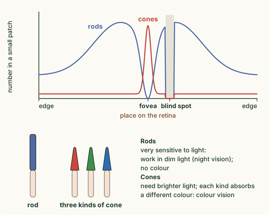
- Rods are more sensitive to light than cones: they respond even to very dim light, so they give night vision. But the rods are all one kind, so rods alone cannot tell colours apart.
- Cones need brighter light. There are three different kinds of cone, and each kind absorbs light of a different colour (roughly red, green and blue light). Together, the three kinds give colour vision.
They are not spread evenly across the human retina. The cones are packed most tightly at the fovea and are few elsewhere. The rods are absent at the fovea, most numerous a little way out from it, and fewer towards the edges of the retina. At the blind spot there are no rods and no cones.
 (enlarge)
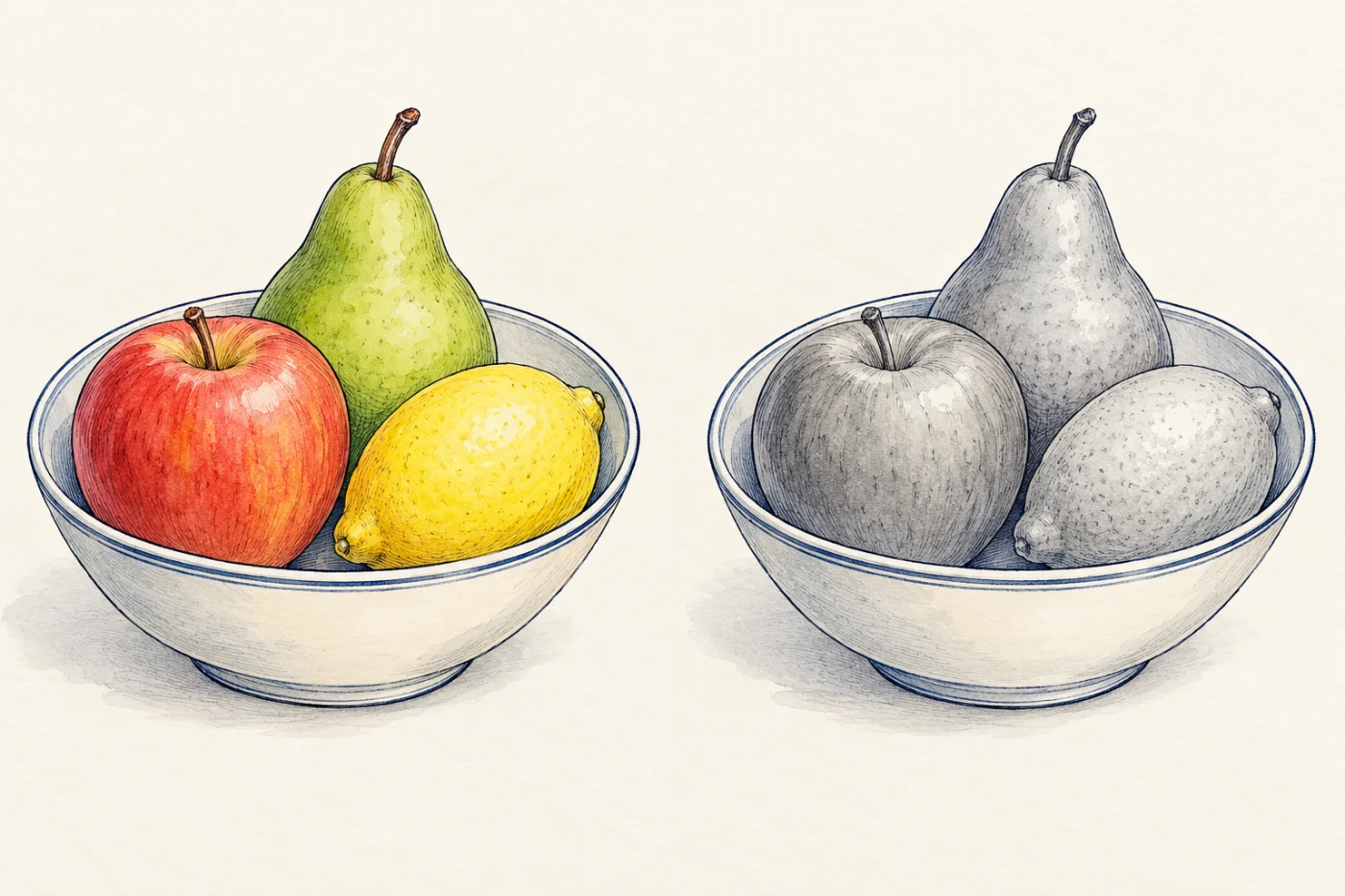
In dim light the cones hardly respond, and you see mostly with your rods. Colours fade, and the scene looks grey, as in the right-hand picture.
Trap: "Rods see colour in the dark." Rods work in dim light, but they give no colour vision. Colour comes from the three kinds of cone, which need brighter light.
The fovea
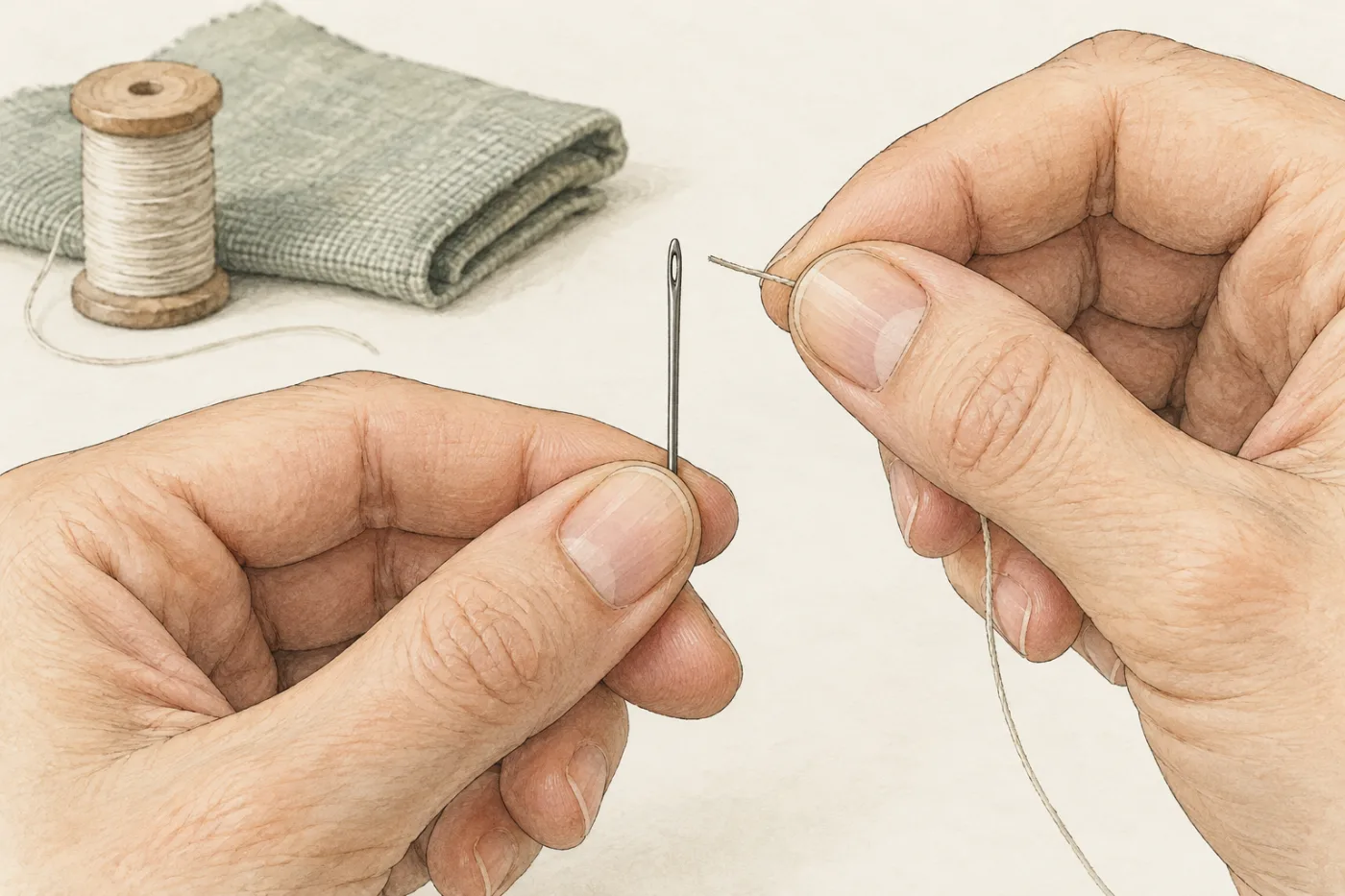
The fovea is a small pit in the retina, at the back of the eye, on the line of sight: when you look straight at something, its image falls on the fovea. The fovea is packed with cones and has no rods. Its function is to give the sharpest, most detailed vision, in colour, in bright light. That is why you look straight at a needle to thread it.
Trap: "The fovea and the blind spot are the same place." The fovea is on the line of sight and is full of cones. The blind spot is to one side, where the optic nerve leaves, and has no receptors at all.
Worked example
From the page to the tree
You look up from a book to a tree far away. Describe the changes in your eye that keep the image sharp.
The tree is distant, so its rays are nearly parallel and need less bending. The ciliary muscles relax, the suspensory ligaments are pulled tight, and the lens is pulled thinner. A thinner lens refracts the light less, so the rays from the tree meet on the retina. Each step is linked: muscle, ligaments, lens shape, refraction.
Worked example
Rays meeting behind the retina
In a model eye, the rays from a near object meet behind the retina. Should the lens become fatter or thinner? Explain.
The rays have not been bent enough: they would meet beyond the retina. The lens must become fatter, so that it refracts the light more and the rays meet on the retina. In a real eye, the ciliary muscles would contract and the suspensory ligaments slacken, letting the lens become fatter.
Worked example
Reading the rods and cones graph
Use the graph of rods and cones to describe where the cones are found in the retina.
The cones are packed most tightly at the fovea, where the graph has a tall, narrow peak, and there are very few anywhere else. The graph also shows that there are no rods at the fovea, and neither rods nor cones at the blind spot. A description names the places on the graph's axis: the fovea, the blind spot, the edges.
Worked example
Grey at night
On a moonlit night, a red car looks grey. Explain why.
The moonlight is dim. The cones, which give colour vision, need brighter light and hardly respond. You see mostly with the rods, which are more sensitive to light and work in dim light but give no colour vision. So the red car looks grey.
Worked example
Looking straight at it
To read small print, you look straight at it rather than out of the corner of your eye. Explain why.
When you look straight at the print, its image falls on the fovea, on the line of sight. The fovea is packed with cones and gives the most detailed vision. Out of the corner of your eye, the image falls further out on the retina, where there are fewer cones, and the detail is lost.
Key idea: (Supplement) Accommodation: for a distant object the ciliary muscles relax, the suspensory ligaments are pulled tight, the lens becomes thinner and refracts light less; for a near object the ciliary muscles contract, the ligaments slacken, the lens becomes fatter and refracts light more. Rods are more sensitive and give night vision; three kinds of cone absorb light of different colours and give colour vision. Cones are packed in the fovea, rods are more numerous away from it, and the blind spot has neither. The fovea lies on the line of sight and gives the most detailed vision.