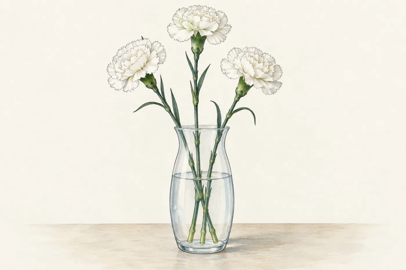
A tall plant takes in water at the bottom, through its roots in the soil, and loses most of it at the top, from its leaves. On a hot day a sunflower moves a lot of water from its roots to its leaves, yet you cannot see any pipes or a pump. Water is colourless, so you cannot watch it move either. But a dye dissolved in the water goes wherever the water goes. Florists use this trick: a white flower with its stem in coloured water slowly turns the colour of the dye. In this part you use a dye to find the route that water takes up a plant.
 (enlarge)
Two transport tissues
A plant has two transport tissues, made of long cells joined end to end that run from the roots, up the stem and into every leaf:
- Xylem carries water and mineral ions from the roots up to the stem, the leaves and the flowers. Its tough walls also help to support the plant, holding it upright.
- Phloem carries sucrose and amino acids, made in the leaves, to the parts of the plant that use or store them.
The xylem and the phloem run side by side in strands called vascular bundles. In a leaf the vascular bundles are the veins that you can see.
Trap: "Phloem carries glucose." Leaves make glucose in photosynthesis, but they change it to sucrose before it is carried. Phloem carries sucrose (and amino acids); xylem carries water and mineral ions.
Tracing water with a dye
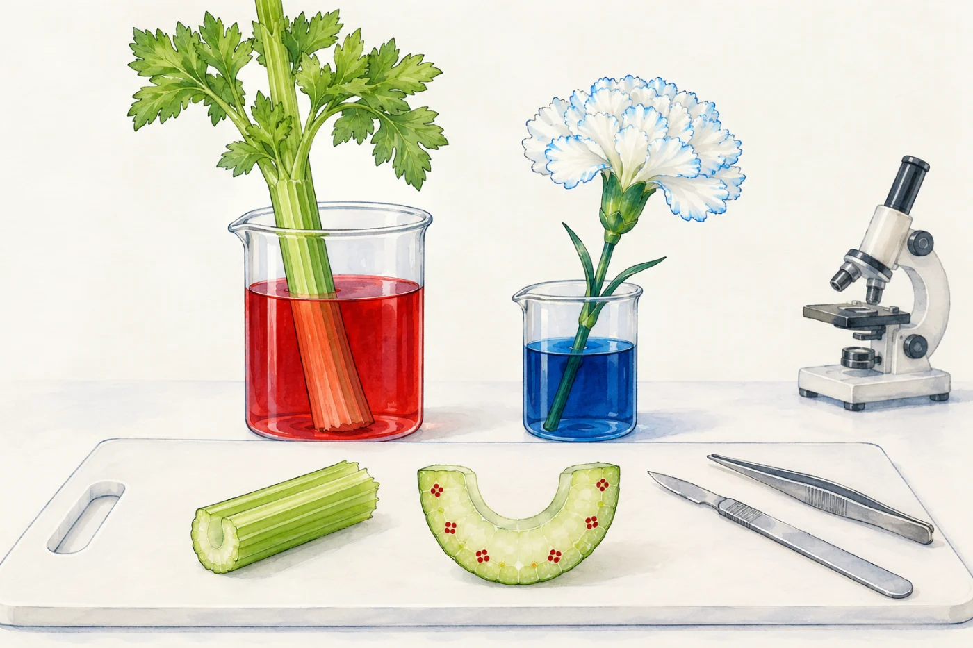
Celery is good for this. The part you eat is a leaf stalk: it joins the leaves to the short stem at the base of the plant. Like a stem, it has vascular bundles running along it, carrying water up to the leaves. The idea is to add a coloured dye to the water: the dye goes where the water goes, and the parts it passes through take up its colour. A part coloured like this is stained. A school method:
- Choose four matched celery stalks: about the same length, each with its leaves still on. Cut about 1 cm off the bottom of each one, so the cut end is fresh.
- Stand stalks A, B and C in a beaker of red dye (red food colouring or eosin solution in water), about 3 cm deep.
- Stand stalk D in a beaker of plain water, the same depth. D is the control: it is treated exactly like the others, except that it has no dye. Any red in A, B or C, but not in D, came from the dye.
- Take out stalk A after 30 minutes, stalk B after 2 hours and stalk C after 6 hours. Take out D at 6 hours too, the longest time. Use a fresh stalk for each time: cutting a stalk into slices destroys it, and a cut stalk put back in the dye would not be a fair comparison.
- Rinse each stalk. On a white tile, use a scalpel to cut a thin slice across it at 4 cm, 10 cm and 16 cm from the cut end. Look at each slice with a hand lens and record where the red is. Look at the leaves of each stalk too.
- Put a very thin slice on a microscope slide, add a drop of water and lower a coverslip onto it. Look at it under the microscope.
 (enlarge)
Celery stalks have no roots: the dye goes in at the cut end. So this experiment shows the route of water through the above-ground parts of a plant. How water gets in through the roots is in part 2.
Predict first: celery stalks will stand in red dye, and later you will cut across them. Where on a cut slice do you think the red will be? All over the cut surface, in separate dots, only in the outer skin, or nowhere? Write your prediction down now.
Below you can try the first steps: setting up the four stalks, then taking out stalk A after 30 minutes and cutting its three slices; the full experiment also cuts B, C and D, looks under the microscope and follows water into the roots. Then read on and compare.
After the experiment: reading the slices
The colours here come from an illustrative model of celery, set up to behave the way real celery usually does. Real stalks vary: the dye climbs faster in a warm, bright room, and more slowly in a cool, dim one. So these times show the pattern, not a speed for water.
| Stalk | In | Time | 4 cm | 10 cm | 16 cm | Leaf veins |
|---|---|---|---|---|---|---|
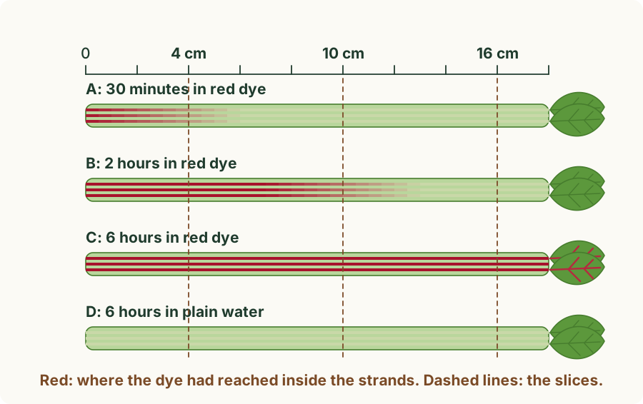
| A | red dye | 30 minutes | faint pink dots | no red | no red | not red |
| B | red dye | 2 hours | dark red dots | red dots | no red | not red |
| C | red dye | 6 hours | dark red dots | dark red dots | dark red dots | red |
| D | plain water | 6 hours | no red | no red | no red | not red |
 (enlarge)
Where the red is. In every slice with red, it was in separate dots in a row near the curved, ridged side, not all over the cut surface. The rest of each slice stayed pale green. The dots are the vascular bundles cut across. So water, carrying the dye, travels up the stalk inside the vascular bundles, not through the whole stalk.
How high it got. The longer a stalk stood in the dye, the higher the red reached. After 30 minutes it was only faint at 4 cm; after 2 hours it reached past 10 cm; after 6 hours it was all the way up, and the dye had gone out of the stalk into the veins of the leaves. Water moves up from the cut end to the leaves.
The control. Stalk D, in plain water for the same 6 hours, had no red at any height. So the red in A, B and C came from the dye, not from the celery itself.
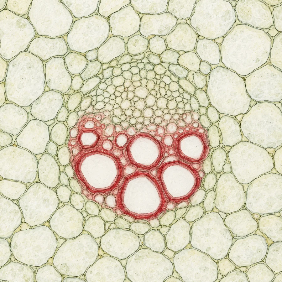
Under the microscope. A thin slice of stalk C shows what is inside one dot. On the outer side of each bundle are groups of small cells: the phloem. On the inner side are wide, empty tubes with thick walls: the xylem vessels. The red was in the walls of the xylem vessels. The phloem stayed pale.
 (enlarge)
So water, with the dye in it, goes up the plant mainly in the xylem.
Trap: "The phloem stayed pale, so phloem carries nothing." The dye only shows where the water went. Phloem carries sucrose and amino acids, which are colourless, so this experiment cannot show them.
Trap: "The red dye soaked through the whole stalk." Only the dots were red. The rest of each slice stayed pale green, because water travels in the vascular bundles.
A white carnation standing in blue dye shows the same thing: after a few hours thin blue lines appear along the veins of its petals. Water carrying the dye has gone up the flower stalk in the xylem and out into the petals.
Worked example
A cabbage leaf in blue dye
A pupil stands the cut end of a white Chinese cabbage leaf in blue food dye for 3 hours, then cuts a slice across its thick white midrib. The slice is white except for a row of small blue dots, and blue lines now run into the thin veins near the base of the leaf. What do the results show about the pathway of water, and what control should be set up?
The dye goes where the water goes. It travelled up the midrib only inside the vascular bundles, the blue dots, not through the whole midrib, and from there it went out into the veins of the leaf. Under a microscope, the blue would be in the walls of the xylem vessels: water moves up the plant in the xylem. After 3 hours the dye had reached only the veins near the base; left longer, it would reach the veins further up. The control is a matched leaf standing in plain water for the same time: if it shows no blue, the colour in the first leaf came from the dye, not from the leaf itself.
Where the xylem and phloem are in a stem
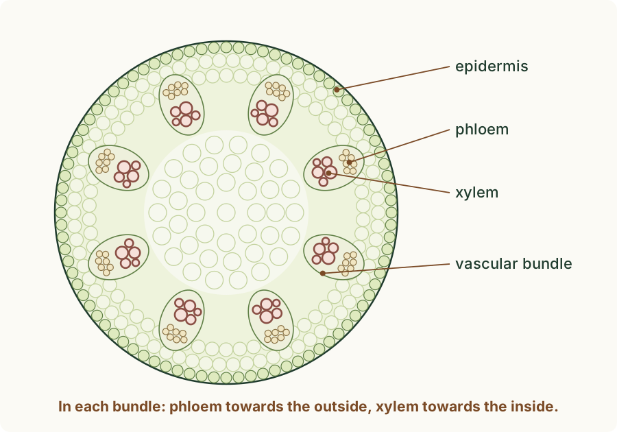
The stem of a non-woody dicotyledonous plant (a flowering plant with a soft stem, not wood, and two seed leaves in each seed), such as a young sunflower or a bean plant, has its vascular bundles in a ring near the outside. In each bundle the phloem is towards the outside of the stem and the xylem is towards the inside. The middle of the stem is filled with large, thin-walled cells.
 (enlarge)
A ring of tough xylem near the outside helps to hold the stem upright. Your celery slices showed the same pattern, in a celery leaf stalk: bundles in a curved row near the ridged side, with phloem on the outer side of each bundle and xylem on the inner side.
Trap: "The xylem is on the outside of the stem." In each bundle of a stem the phloem is on the outer side and the xylem on the inner side.
Worked example
A tall sunflower stem
A young sunflower grows 2 m tall, with a heavy flower head in which hundreds of seeds are filling with food. A thin slice is cut across its soft stem. Where in the slice are the xylem and the phloem, and what does each tissue do for the plant?
The vascular bundles form a ring near the outside of the stem. In each bundle the phloem is on the outer side and the xylem on the inner side, towards the middle. The xylem carries water and mineral ions from the roots up to the leaves and the flower head, and its tough walls help to support the tall stem: the ring of xylem near the outside helps to hold it upright. The phloem carries sucrose and amino acids, made in the leaves, to the parts that use or store them, such as the filling seeds.
Supplement: xylem vessels
This section is Supplement content (syllabus 8.1.3).
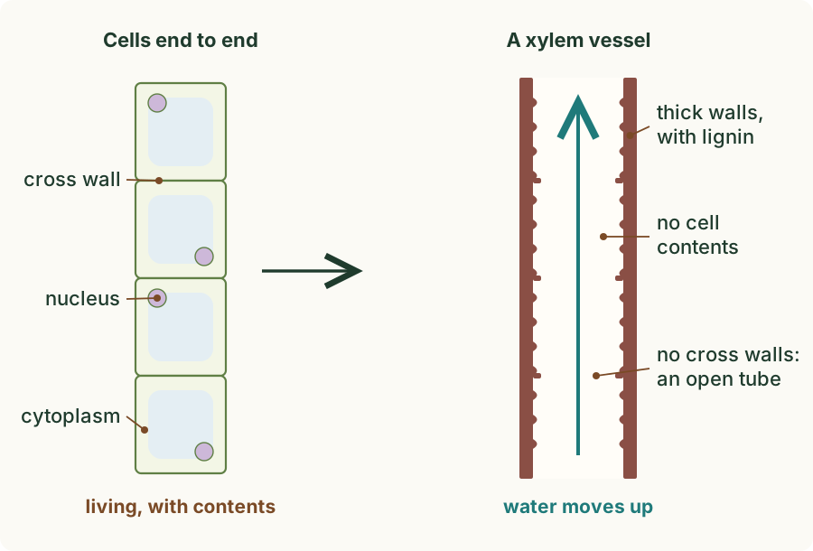
A xylem vessel forms from a column of cells joined end to end. As it develops, three things happen, and each one suits it to carrying water:
- Its walls become thick, strengthened with lignin. The strong walls keep the tube open, so it does not collapse, and they help to support the plant.
- The cells lose their contents: a finished vessel has no cell contents, no cytoplasm and no nucleus, so nothing inside blocks the water.
- The cross walls between the cells break down. With no cross walls, the cells make one long, continuous tube, and water moves up it freely. Vessels join up with one another, so the xylem gives water an unbroken path from the roots to the leaves.
 (enlarge)
Trap: "Living cells inside the xylem pump the water up." A xylem vessel has no cell contents: it is an empty tube. Nothing inside it pushes the water.
Worked example
A model vessel made of cans
Supplement. A teacher makes a model of a xylem vessel: she cuts both ends out of five empty tin cans and tapes them end to end into one long tube. Explain how each part of the model matches the structure of a real xylem vessel, and how each feature helps it carry water.
Each can stands for one cell, and taping the cans end to end matches the column of cells that a vessel forms from. Cutting out the ends matches the cross walls breaking down: with no cross walls, the cells make one long, continuous tube, and water moves up it freely. The cans are empty, like a finished vessel, which has no cell contents, no cytoplasm and no nucleus, so nothing inside blocks the water. The stiff metal stands for the thick walls strengthened with lignin, which keep the tube open so that it does not collapse, and help to support the plant.
Key idea: Xylem carries water and mineral ions up the plant and helps to support it; phloem carries sucrose and amino acids. They run together in vascular bundles. A dye in the water travels up a celery stalk in the vascular bundles, mainly in the xylem, and reaches higher the longer the stalk stands in it, until it reaches the leaf veins; a matched stalk in plain water is the control. In a non-woody dicotyledonous stem the bundles form a ring near the outside, with phloem towards the outside and xylem towards the inside. (Supplement) Xylem vessels have thick walls with lignin, no cell contents and no cross walls, making a long, continuous tube for water.