At rest you breathe about six litres of air in and out every minute, and in part 1 you found that the air comes back out with less oxygen and more carbon dioxide in it. So somewhere inside your chest, gases are passing between the air and your blood. In this part you follow the air down to the place where that happens, and find out why it happens so fast there.
The breathing system
 (enlarge)
Follow a breath in, from your mouth or nose to the end of the line.
 (enlarge)
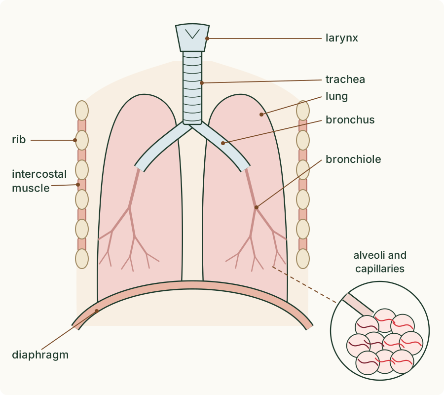
- The air passes the larynx, the voice box, at the top of the windpipe.
- It goes down the trachea, the windpipe, a tube held open by rings of cartilage.
- The trachea divides into two bronchi (one bronchus to each lung).
- Inside each lung, the bronchi branch again and again into thousands of narrower tubes, the bronchioles.
- Each bronchiole ends in a cluster of tiny air sacs, the alveoli (one alveolus). Every alveolus is wrapped in a net of blood capillaries, the narrowest blood vessels.
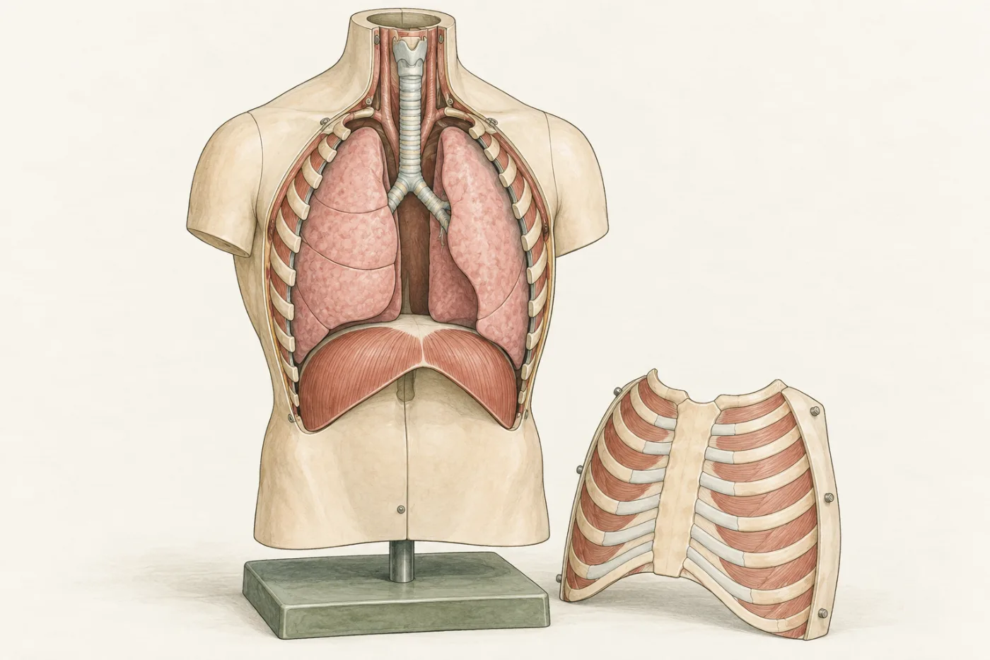
The lungs sit inside the chest, or thorax, protected by the ribs. Between neighbouring ribs are the intercostal muscles ("intercostal" means "between the ribs"). Under the lungs lies the diaphragm, a dome-shaped sheet of muscle that separates the thorax from the abdomen below. The ribs, the intercostal muscles and the diaphragm move the air in and out, as part 3 shows.
Trap: "The lungs are two hollow bags." Each lung is spongy: it is filled with millions of alveoli and the tubes that lead to them, with blood vessels running through it all.
Where the gases cross
In the experiment, look at one alveolus and the capillary beside it in a model, then take a breath in.
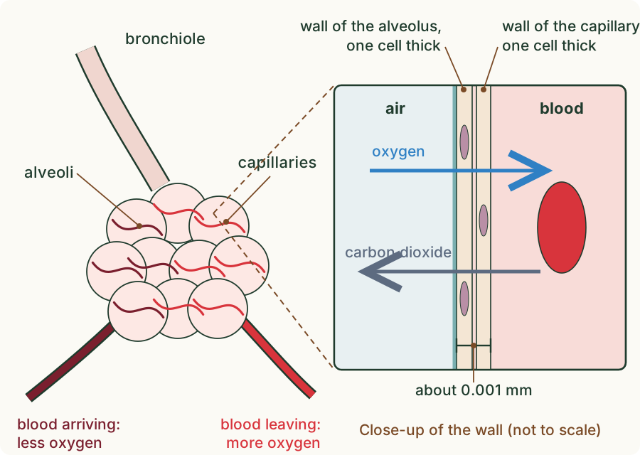
Oxygen crosses from the air in the alveolus into the blood, and carbon dioxide crosses the other way, both by diffusion. A surface where gases cross between an organism and its surroundings is called a gas exchange surface, and in humans it is the lining of the alveoli. Diffusion is fast across it because it has four features.
 (enlarge)
- A large surface area. The two lungs hold about 300 million alveoli. Their walls together cover about 70 m², roughly the floor of a classroom, so a great deal of gas can cross at once.
- A thin surface. The wall of an alveolus and the wall of the capillary next to it are each only one cell thick, so together they are about a thousandth of a millimetre across. Gases have only a very short distance to diffuse.
- A good blood supply. A dense network of capillaries covers the alveoli. The blood carries oxygen away as soon as it arrives and brings more carbon dioxide, so the blood next to the air always has less oxygen and more carbon dioxide than the air. That difference in concentration keeps diffusion going.
- Good ventilation with air. Breathing replaces the air in the alveoli again and again, so the air there stays high in oxygen and low in carbon dioxide, and the difference with the blood never runs out.
The lining is also moist, and the gases dissolve in that thin layer of water as they cross; that is where the water vapour in expired air comes from.
Trap: "Alveoli are cells." An alveolus is a tiny air sac, about a fifth of a millimetre across. Its wall is made of very thin cells.
(Supplement) Keeping the trachea open
 (enlarge)
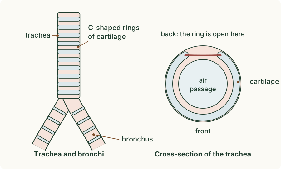
The wall of the trachea contains C-shaped rings of cartilage, a firm but slightly bendy tissue (you can feel it in the tip of your nose). When you breathe in, the air pressure inside the airways falls, and a soft tube would be squashed flat. The cartilage holds the trachea open, so air can always pass. The bronchi have cartilage too.
(Supplement) Two sets of intercostal muscles
 (enlarge)
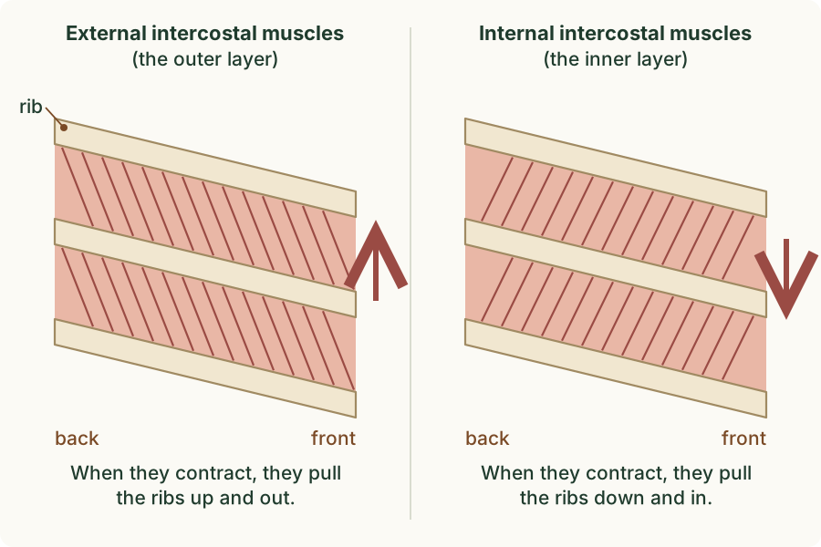
There are two layers of intercostal muscles between each pair of ribs. The external intercostal muscles are on the outside; the internal intercostal muscles lie inside them, and their fibres slant the other way. A muscle can only pull when it contracts, so the two layers work against each other: the external intercostals pull the ribs up and out, and the internal intercostals pull them down and in. When one layer contracts, the other relaxes; a pair of muscles that work like this is called antagonistic. Part 3 shows when each layer works.
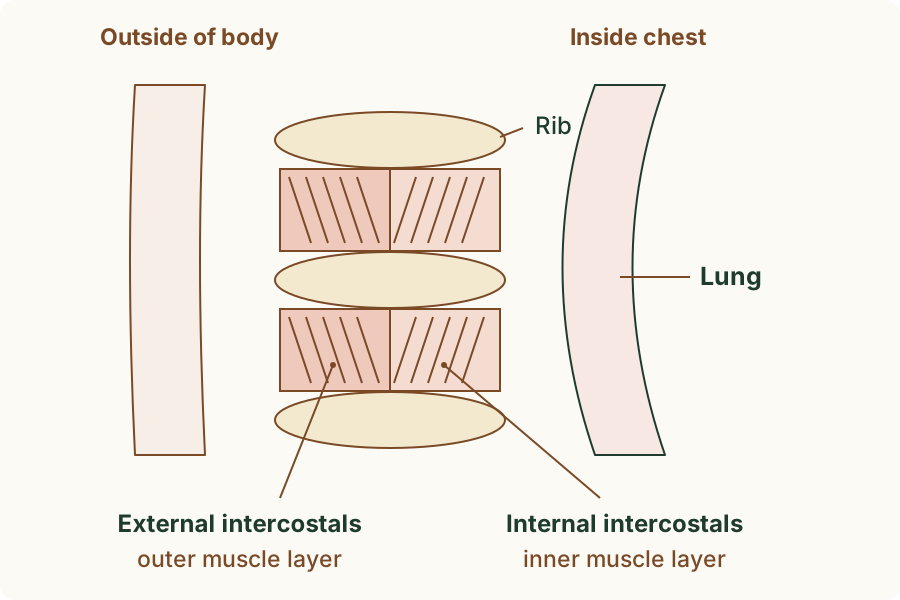
In a section through the chest wall you can tell the two layers apart by where they are: the external intercostals are the layer nearer the outside of the body, and the internal intercostals the layer nearer the lung.
 (enlarge)
(Supplement) Keeping the air clean
Air carries dust, pollen and pathogens (microorganisms that cause disease, such as bacteria and viruses). If they reached the alveoli, they could damage the thin gas exchange surface or cause an infection there. The trachea and bronchi are lined with cells that stop most of them on the way.
 (enlarge)
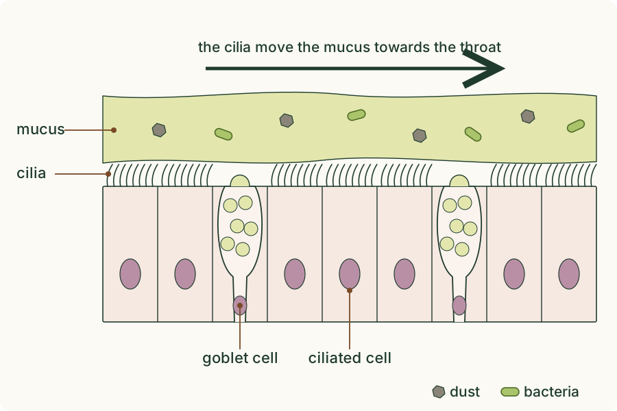
- Goblet cells make mucus, a sticky liquid that spreads over the lining. Particles and pathogens stick to it and are trapped.
- Ciliated cells carry cilia, tiny hair-like structures that beat together like rowers' oars. They sweep the mucus, with what it has trapped, up the trachea to the throat, where it is swallowed. Most pathogens are then killed by the acid in the stomach.
Trap: "Cilia trap the dust." The mucus traps it; the cilia move the mucus along.
Worked example
The path of a breath
Name, in order, the parts that air passes through from the larynx to the blood.
Larynx → trachea → bronchus → bronchioles → alveolus; then oxygen diffuses through the wall of the alveolus and the wall of a capillary into the blood. A common slip is to put the bronchioles before the bronchi: the bronchi are the two large branches of the trachea, and the bronchioles are the much smaller branches inside the lungs.
Worked example
Many small sacs
Explain why lungs made of millions of tiny alveoli are better for gas exchange than two large hollow bags holding the same volume of air.
Splitting the same volume into millions of tiny sacs gives a far larger surface area, about 70 m² in human lungs, while two large bags of the same volume would have well under 1 m². More surface means more oxygen and carbon dioxide can diffuse at once. The tiny sacs also keep every bit of air close to a thin wall with a capillary behind it, so the diffusion distance stays short.
Worked example
Floppy airways
(Supplement) A rubber tube with soft walls is attached to a pump that sucks air through it. The tube flattens and the air stops. Explain how the trachea avoids this problem.
When the pump sucks, the pressure inside the soft tube falls below the pressure outside, so the walls are pushed in and the tube collapses. When you breathe in, the pressure in the trachea also falls, but its wall has C-shaped rings of cartilage, which are firm enough to keep the trachea open, so air keeps flowing to the lungs.
Worked example
A dusty workplace
(Supplement) A worker sweeps a dusty floor all day. Explain how the lining of the trachea stops most of the dust and bacteria in the air from reaching the alveoli.
Goblet cells in the lining make mucus, which traps the dust particles and bacteria. Ciliated cells have cilia that beat and move the mucus up the trachea to the throat, where it is swallowed, so the trapped particles and pathogens are carried away from the lungs. Very fine dust that gets past this defence can still reach the alveoli, which is why dust that is breathed in for years can damage the lungs.
Key idea: Air passes the larynx, then down the trachea, the bronchi and the bronchioles to the alveoli, which are wrapped in capillaries; the lungs lie inside the ribs, with intercostal muscles between the ribs and the diaphragm below. The alveoli are a gas exchange surface: a large surface area, a thin surface, a good blood supply and good ventilation with air make diffusion fast. (Supplement) External and internal intercostal muscles pull the ribs in opposite directions; cartilage keeps the trachea open; mucus from goblet cells traps pathogens and particles, and the cilia of ciliated cells sweep it up to the throat.