The lungs have no muscles that can pull them open, yet they fill with air about twelve times a minute while you sit still, and much more often when you run. In this part you find out what moves the air in and out, using a model of the chest, and then measure how breathing changes with exercise from a volunteer's breathing traces.
Moving air with a model
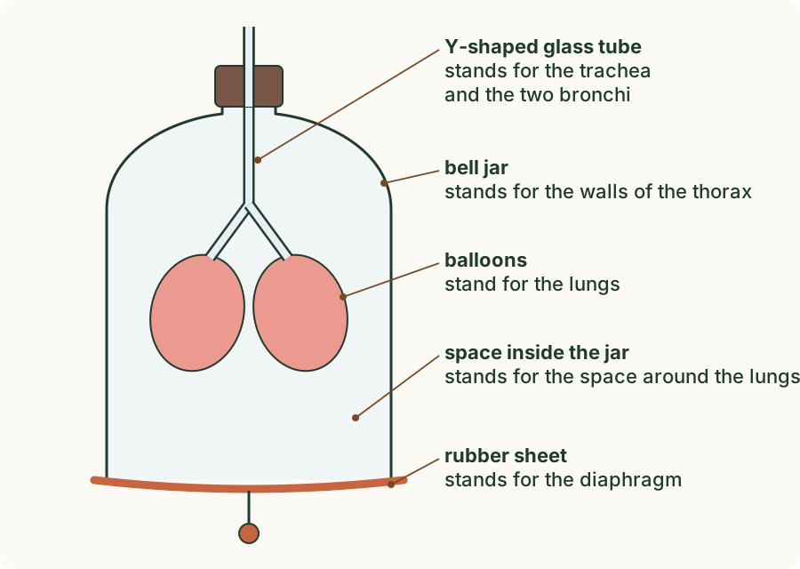
The bell-jar model is a simple model of the chest. Each part stands for a part of the body.
 (enlarge)
| Part of the model | What it stands for |
|---|---|
| glass jar | the walls of the thorax (the ribs) |
| rubber sheet | the diaphragm |
| Y-shaped tube | the trachea and the two bronchi |
| balloons | the lungs |
| the space inside the jar, around the balloons | the space inside the thorax, around the lungs |
In the experiment, predict what the balloons will do when you pull the rubber sheet down, then try it.
(Supplement) What the model showed
When you pulled the sheet down, the balloons filled with air. When you pushed it back up, they emptied. Nothing pushed air into the tube, so how did it get in?
- Pulling the sheet down makes the volume inside the jar larger.
- The same air now has more room, so its pressure falls. The pressure inside the jar drops below the pressure of the air outside, the atmospheric pressure.
- Air always moves from a higher pressure to a lower pressure, so air from outside flows in through the tube and fills the balloons.
- Pushing the sheet up makes the volume smaller, the pressure inside rises above atmospheric pressure, and air is pushed out.
So the order is always volume, then pressure, then air. The balloons fill because the space around them gets bigger, not because they suck air in.
Trap: "The lungs suck air in, and that pushes the chest out." It happens the other way round: muscles make the chest bigger first, the pressure inside falls, and then air flows in.
(Supplement) Where the model falls short
A model leaves things out, and this one leaves out a lot.
- The jar's walls are rigid glass, but the real ribs move up and out, which makes the thorax larger too. The model has no ribs that move and no intercostal muscles.
- The rubber sheet is flat and is pulled by hand. The real diaphragm is a dome of muscle that flattens when it contracts.
- The balloons are hollow bags. Real lungs are spongy, filled with alveoli.
- In the jar there is a lot of air around the balloons. In the body, the lungs fill almost all of the thorax, with only a very thin layer of fluid between them and the chest wall.
(Supplement) Breathing in
 (enlarge)
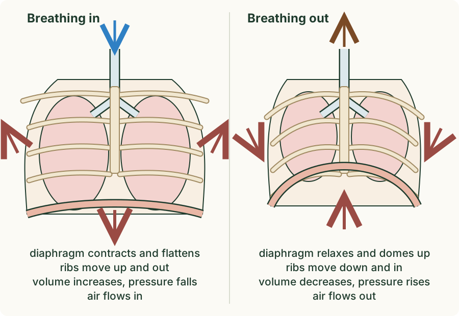
Breathing in is called inspiration. It takes muscle work.
- The diaphragm contracts and flattens, moving down.
- The external intercostal muscles contract and the internal intercostal muscles relax, so the ribs move up and out.
- Both movements increase the volume of the thorax.
- The pressure inside the thorax falls below atmospheric pressure.
- Air flows in through the trachea into the lungs.
(Supplement) Breathing out
Breathing out is called expiration.
- At rest, the diaphragm relaxes and is pushed back up into its dome, and the external intercostal muscles relax, so the ribs move down and in. The volume of the thorax decreases, the pressure inside rises above atmospheric pressure, and air is pushed out. The stretched lungs also spring back, like a balloon letting go.
- When you breathe out hard, during exercise or a cough, the internal intercostal muscles contract and pull the ribs down and in further and faster, and the muscles of the abdomen push the diaphragm up. More air is pushed out, and faster. This is forced expiration.
So the external and internal intercostal muscles are antagonistic: they pull the ribs in opposite directions, and when one set contracts, the other relaxes.
Breathing during exercise
When you exercise, you breathe faster and more deeply. Two measurements describe this.
- The rate of breathing is the number of breaths per minute.
- The depth of breathing is the volume of air breathed in (or out) in one breath.
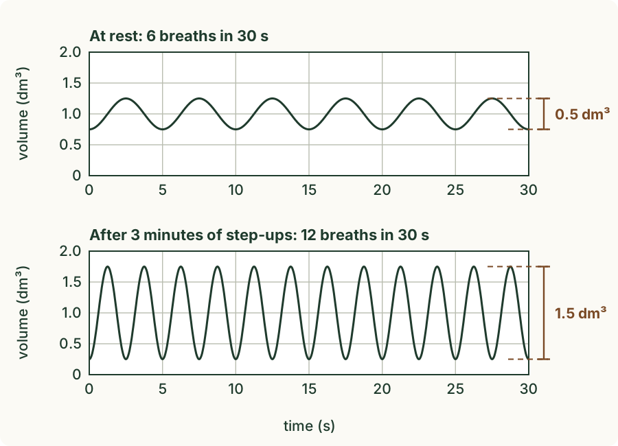
A spirometer (a machine that measures the volume of air you breathe in and out) with a sensor and a data logger (a device that records the readings and draws them as a graph) record a volunteer's breathing as a trace: a line that goes up with each breath in and down with each breath out. Each peak is one breath, and the height of a peak above the dip before it is the depth of that breath. Volumes are in dm³ (cubic decimetres); 1 dm³ is the same as 1 litre, or 1000 cm³.
Counting for a whole minute takes a long time, so you count the breaths in 30 seconds and multiply by 2, because a minute (60 s) holds two lots of 30 s (60 ÷ 30 = 2). If you counted for 15 s, you would multiply by 4 (60 ÷ 15 = 4).
 (enlarge)
What the traces showed
| Breaths in 30 s | Rate (breaths per minute) | Depth (dm³) | Air per minute (dm³) | |
|---|---|---|---|---|
| At rest | 6 | 6 × 2 = 12 | 0.5 | 12 × 0.5 = 6 |
| After 3 minutes of step-ups | 12 | 12 × 2 = 24 | 1.5 | 24 × 1.5 = 36 |
After exercise, the rate doubled (from 12 to 24 breaths per minute) and the depth tripled (from 0.5 dm³ to 1.5 dm³). Together they move six times as much air through the lungs each minute (36 ÷ 6 = 6). So physical activity increases both the rate and the depth of breathing. A few minutes after the exercise stops, both fall back to the resting values.
To trust a result like this, the investigation keeps everything else the same: the same volunteer, the same exercise for the same time, the counts taken the same way (30 s, straight after the exercise), and repeats with several people to find an average.
(Supplement) Why breathing speeds up
 (enlarge)
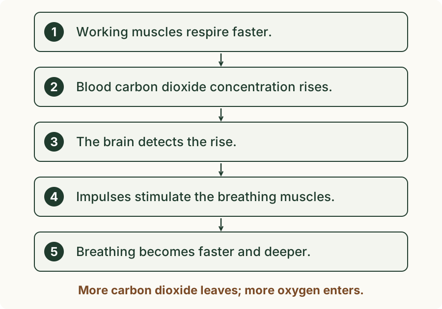
- During exercise, the muscles contract more, so they need more energy and respire faster.
- Aerobic respiration makes carbon dioxide, so the concentration of carbon dioxide in the blood rises.
- The brain detects the higher carbon dioxide concentration in the blood.
- The brain sends nerve impulses to the diaphragm and the intercostal muscles, so they contract more often and more strongly.
- Breathing becomes faster and deeper. More carbon dioxide is breathed out, and more oxygen reaches the blood for the muscles' respiration.
Trap: "Breathing speeds up because the muscles run out of oxygen." The signal the brain detects is the rise in carbon dioxide in the blood. The extra oxygen is a result of faster, deeper breathing, not its trigger.
Worked example
Reading the model
(Supplement) In the bell-jar model, explain why the balloons fill with air when the rubber sheet is pulled down, and give one way in which the model is not like a real chest.
Pulling the sheet down increases the volume inside the jar, so the pressure inside falls below atmospheric pressure. Air flows from the higher pressure outside, through the Y-shaped tube, into the balloons, which fill. One difference: the jar's walls cannot move, but real ribs move up and out, moved by the intercostal muscles, which the model does not have. (Other answers: the diaphragm is a dome of muscle, not a flat sheet pulled by hand; lungs are spongy, not hollow.)
Worked example
One breath in
(Supplement) Describe how the muscles of the thorax cause air to enter the lungs.
The diaphragm contracts and flattens. The external intercostal muscles contract (the internal ones relax), so the ribs move up and out. The volume of the thorax increases, so the pressure inside falls below atmospheric pressure, and air flows in through the trachea to the lungs. A good answer keeps the order: muscles, then volume, then pressure, then air.
Worked example
A breathing trace
A spirometer trace shows that a pupil breathes 4 times in 15 seconds, and each breath has a depth of 0.8 dm³. Find the pupil's rate of breathing and the volume of air they breathe each minute.
A minute holds four lots of 15 s (60 ÷ 15 = 4), so the rate is 4 × 4 = 16 breaths per minute. Each breath moves 0.8 dm³, so the volume per minute is 16 × 0.8 = 12.8 dm³ per minute. Check the units: breaths per minute × dm³ per breath gives dm³ per minute.
Worked example
Why breathing speeds up
(Supplement) A runner's breathing becomes faster and deeper during a race. Explain why.
The leg muscles contract more, so they respire faster and make more carbon dioxide, which raises the carbon dioxide concentration in the blood. The brain detects this rise and sends nerve impulses to the diaphragm and intercostal muscles, which contract more often and more strongly. Breathing becomes faster and deeper, so the extra carbon dioxide is removed and more oxygen reaches the blood for respiration.
Key idea: Physical activity increases both the rate (breaths per minute) and the depth (volume per breath) of breathing; the rate is found by counting breaths for 30 s and multiplying by 2. (Supplement) Breathing in: the diaphragm contracts and flattens and the external intercostal muscles pull the ribs up and out, so the volume of the thorax increases, the pressure falls below atmospheric pressure and air flows in. Breathing out: the muscles relax (and in forced expiration the internal intercostals pull the ribs down and in), the volume decreases, the pressure rises and air flows out. During exercise, respiring muscles raise the carbon dioxide concentration in the blood; the brain detects it and makes breathing faster and deeper.