This part is Supplement content (syllabus 7.5.4 and 7.5.5).
At ×100 the villi were a row of fingers along the inner edge of the ileum. A large surface is only half the story: once a glucose molecule has crossed the lining, something has to carry it away, or the villus would soon be full. In this part you look inside one villus at ×400 and follow where the absorbed molecules go.
A closer look
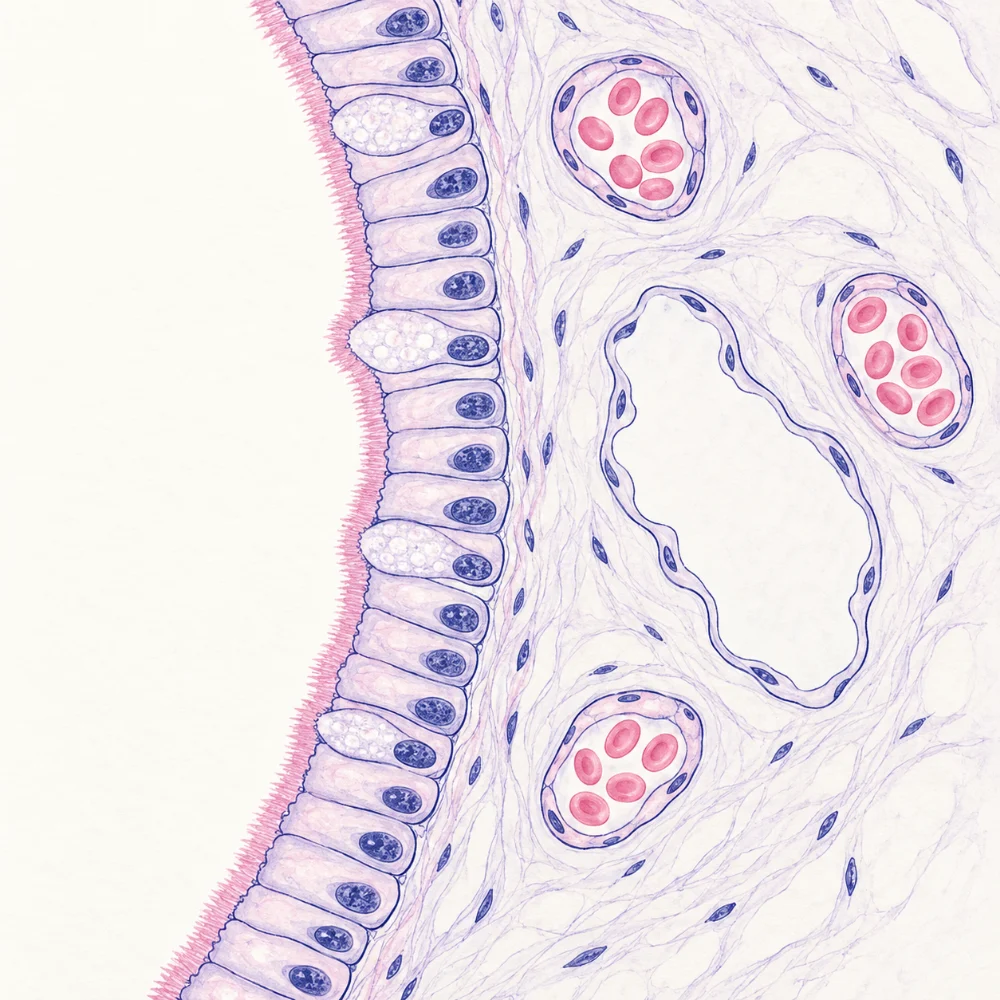
Turn the nosepiece to the ×40 objective: with the ×10 eyepiece that is ×400. The field of view is now only about 0.45 mm across, so you see part of one villus, not a whole one. Look for the lining, and for anything inside the villus that could carry molecules away. Then run the experiment below before you read on.
What the ×400 field showed
 (enlarge)
The lining is a single row of cells: the epithelium is one cell thick. The fuzzy band along its outer edge is where the microvilli are, too small to see one by one at ×400. (The pale, rounded cells make mucus, which protects the lining.) Behind the lining, small vessels full of red blood cells are capillaries, cut across. The larger space with no blood cells in it is the lacteal.
The structure of a villus
A section shows only what the slice happened to cut through. Putting many sections together gives the whole villus:
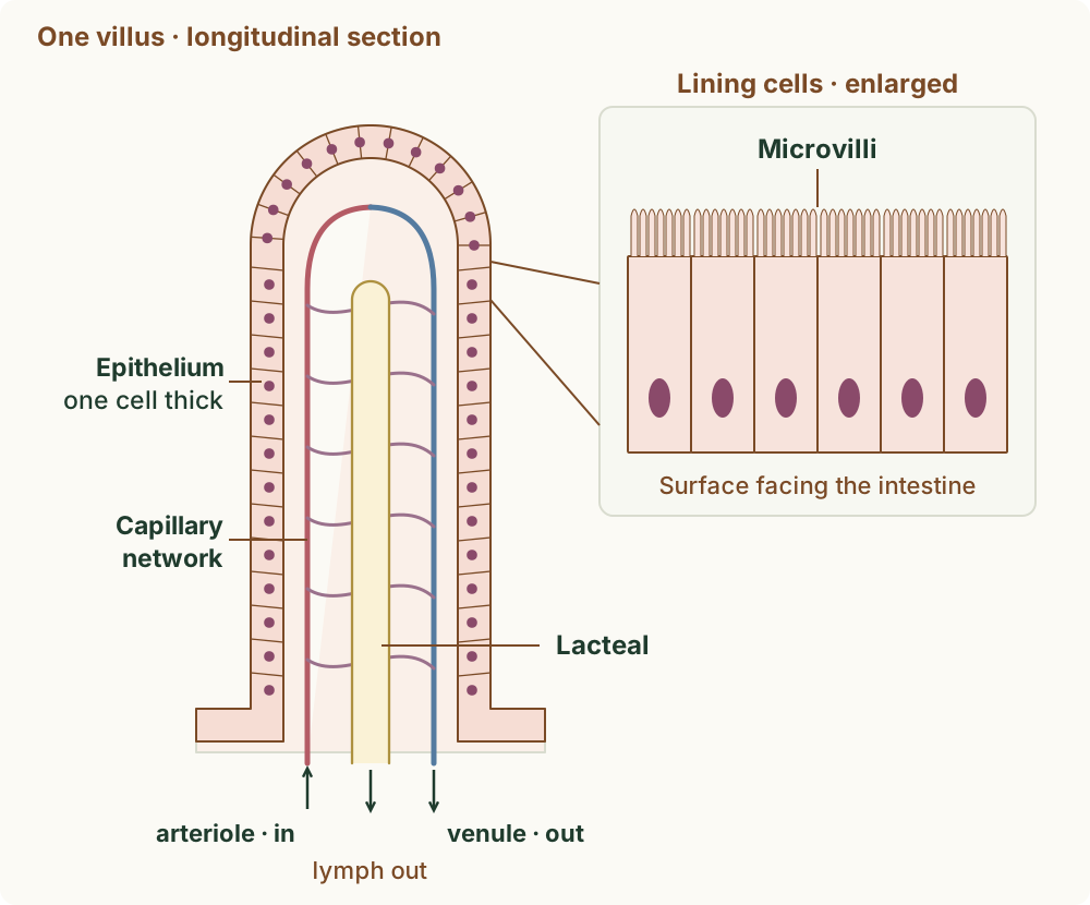
- the epithelium, one cell thick, covers its surface. Each epithelial cell has microvilli on the side facing the food;
- just under the epithelium is a network of capillaries. Blood comes in from a small artery (an arteriole) at the base, flows through the network and leaves along a small vein (a venule);
- in the middle is the lacteal, a lymph vessel closed at its tip. It carries lymph, a pale fluid, out of the villus at its base.
 (enlarge)
The drawing is a teaching reconstruction: it puts together the parts that different sections show, and the microvilli are in an inset magnified much more. Because the epithelium is only one cell thick, absorbed molecules have only a short distance to travel from the food to the blood.
Trap: "The lining of a villus is thick, to protect it." The epithelium is one cell thick. A thin lining gives absorbed molecules a short distance to travel to the capillaries and the lacteal.
Worked example
Describing a villus
Supplement. A student draws a villus as a finger shape with a lining three cells thick and nothing inside it. Describe four things a correct drawing should show.
A correct drawing shows: (1) an epithelium one cell thick covering the villus; (2) microvilli on the epithelial cells, on the side facing the space inside the intestine (in a magnified inset, since they are too small to draw to scale); (3) a network of capillaries just under the epithelium, joined to an arteriole and a venule at the base; (4) a lacteal in the middle of the villus, closed at its tip.
Capillaries and lacteals
The two kinds of vessel take different molecules away.
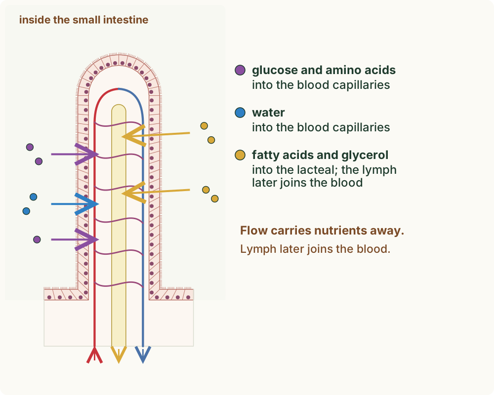
- Capillaries absorb glucose and amino acids, and also water, into the blood. The blood flowing through the capillaries carries them away from the villus, to the rest of the body.
- The lacteal absorbs fatty acids and glycerol, the products of fat digestion, into the lymph. The lymph flows out of the villus into larger lymph vessels, and later empties into the blood near the heart.
 (enlarge)
Blood flows through the capillaries all the time, so absorbed molecules are carried away as fast as they arrive and the villus never fills up: absorption can go on for as long as there is digested food in the intestine.
Trap: "The lacteal carries blood." The lacteal is a lymph vessel: it carries lymph, with the fatty acids and glycerol, not blood. Glucose and amino acids go into the blood capillaries.
Worked example
A fatty meal
Supplement. A meal of chips fried in oil is digested in the small intestine. Name the molecules made from the oil and from the starch in the potato, and describe the route each takes into a villus.
The oil is a fat: lipase breaks it into fatty acids and glycerol. These pass through the epithelium of a villus into the lacteal, and are carried away in the lymph, which later joins the blood. The starch is broken down to glucose, which passes through the epithelium into the capillaries and is carried away in the blood.
Worked example
More glucose leaving
Supplement. Blood is sampled from an arteriole going into the wall of the small intestine and from a venule coming out of it, an hour after a meal. The blood in the venule contains much more glucose. Explain this, using the structure of the villi.
Starch in the meal has been digested to glucose in the small intestine. Glucose molecules pass through the epithelium of the villi, which is one cell thick, into the capillary network just under it. Blood flowing through the capillaries picks up the glucose and leaves along the venule, so the venule's blood has more glucose than the arteriole's. The huge surface of the villi and microvilli means a lot of glucose is absorbed in the time the blood takes to pass through.
So much, so quickly
The small intestine absorbs so much so quickly because its lining has a very large surface, folded into villi with microvilli on every epithelial cell; the epithelium is one cell thick, so the distance to the blood is short; and blood and lymph keep flowing, carrying absorbed molecules away through the capillaries and the lacteals.
Key idea: (Supplement) A villus is a finger-like projection of the lining of the small intestine. It is covered by an epithelium one cell thick, with microvilli on its cells; it has a network of capillaries under the epithelium and a lacteal in the middle. The capillaries absorb glucose and amino acids into the blood, which carries them away; the lacteals absorb fatty acids and glycerol into the lymph, which later joins the blood.