This part is Supplement content (syllabus 7.5.3).
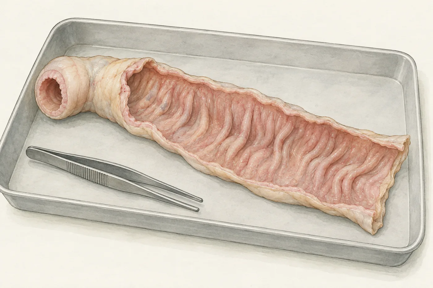
A piece of small intestine from an animal, cut open along its length, is not smooth inside. Its lining is folded into ridges, and under a hand lens the ridges look like velvet. The small intestine has to absorb a whole day's digested food, and most of 9 litres of water, in the few hours that food spends passing through it. In this part you look at its lining under the microscope and measure how much the folding adds.
 (enlarge)
A folded lining
The surface that the digested food touches, on the inside of the small intestine, is its internal surface area. Absorption happens across this surface: the larger it is, the more molecules can pass through it in the same time.
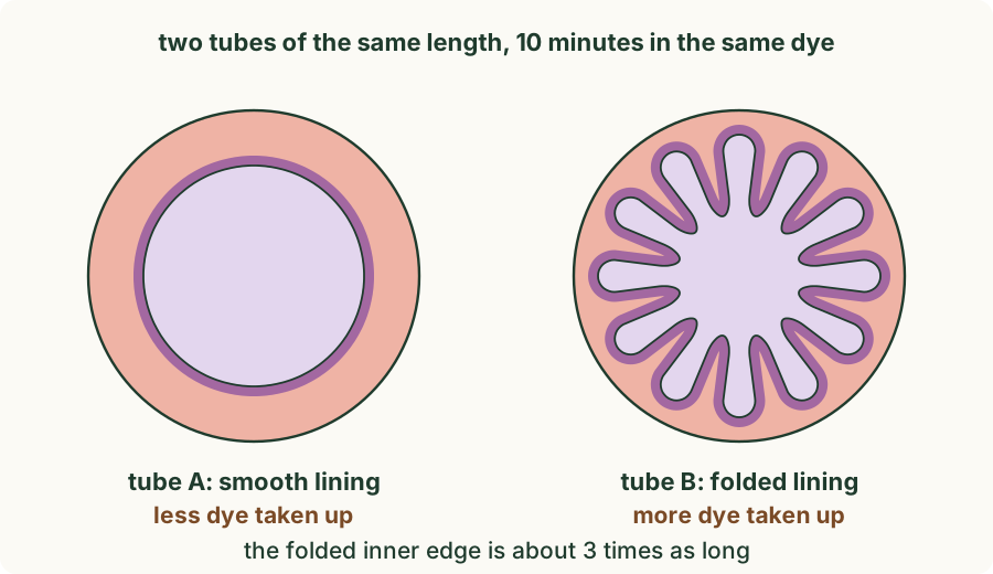
Two tubes of the same length show the idea. Their linings take up a purple dye. Tube A's lining is smooth; tube B's is folded into twelve folds that stick out into the middle.
 (enlarge)
Worked example
Smooth and folded tubes
The two tubes in the drawing are the same length and spend the same time in the same dye. Explain why tube B takes up more dye than tube A.
Both tubes are the same length, so any difference comes from the lining. Tube B's lining is folded: its inner edge is about 3 times as long as tube A's, so along the whole tube its inner surface is larger. More of the lining touches the dye at the same time, so more dye is taken up in the same 10 minutes. The folding changes the surface, not the length of the tube.
Measuring the folded edge
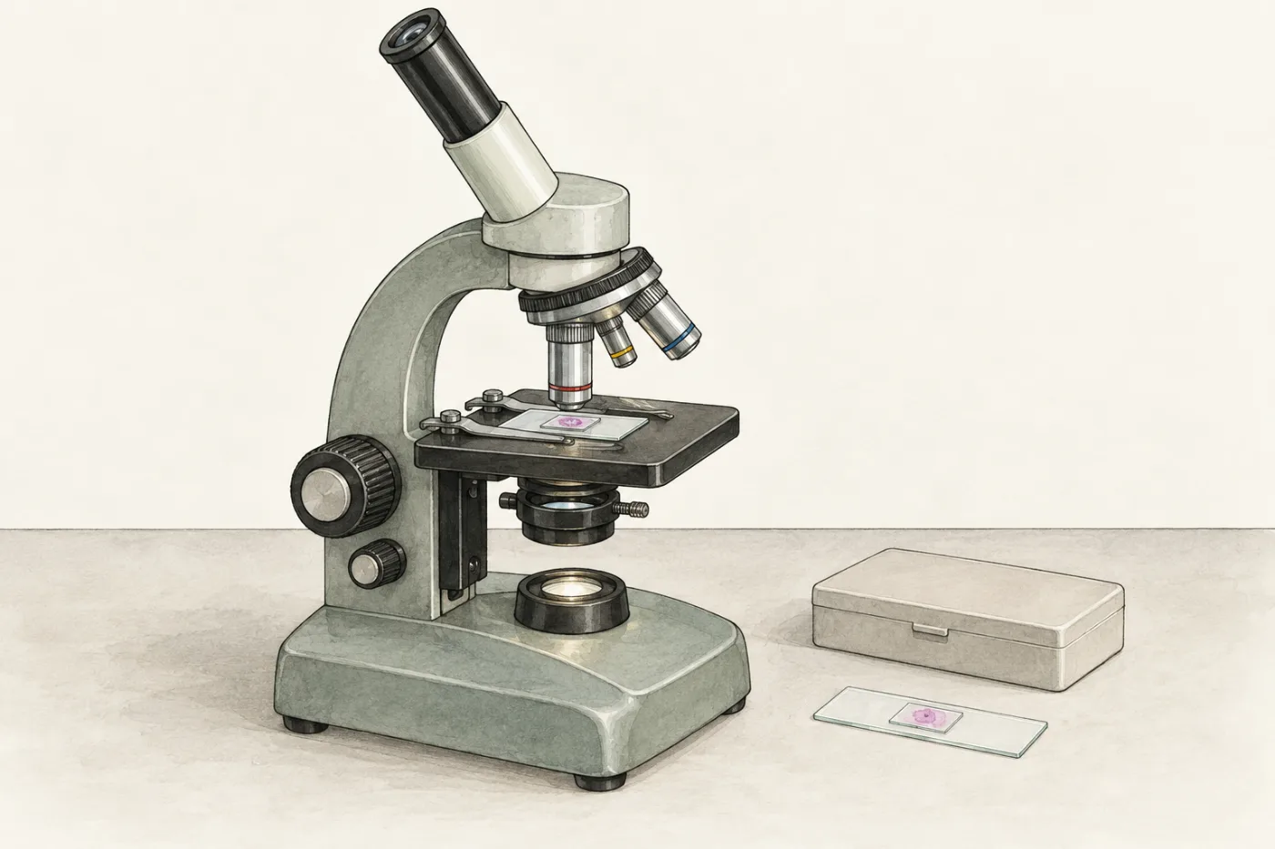
The real lining is folded far more finely. A prepared slide is a very thin slice of tissue, stained so that its parts show up, sealed under a cover slip. The slice on this slide was cut across the wall of the ileum. Under the microscope you will see its inner edge, and you will lay a string along the edge to measure how much longer the folded edge is than the straight distance between its ends.
Predict first: if you straightened out the folded edge of a stretch of lining, how many times longer would it be than the straight distance between its ends? About the same, about twice, four or five times, or about a hundred times? Make your prediction, then run the experiment below before you read on.
 (enlarge)
What the string showed
At ×100 the inner edge of the ileum is a row of fingers, each about half a millimetre long. These are villi (one is a villus): finger-like projections of the lining, millions of them, sticking out into the space inside the small intestine.
| Stretch of the edge | Straight length | String along the edge | Times longer |
|---|---|---|---|
| A, three whole villi | 1.05 mm | 4.31 mm | 4.1 |
| B, four whole villi | 1.22 mm | 5.69 mm | 4.7 |
These are the lengths along the whole edge. A string pinned only at the tips of the villi and the bottoms of the gaps cuts the round tips a little short, so it gives a slightly smaller ratio: in the experiment, where both strings are pinned that way, stretch A gives about 3.8 and stretch B 4.3. Either way, along the same straight distance the folded edge is about four times longer: the mean of the two stretches is about 4.4 along the whole edge, and about 4.1 with pins only at the tips and gaps. The right-hand edge of the first field was no good for measuring: there the villi were cut across at a slant, and the edge broke up into separate islands.
The ratio comes from one thin slice. It compares two lengths, not two areas: real villi are fingers, so they add surface all round, not just along one edge. And it says nothing about how fast molecules cross the lining. What it does show is that the villi make the surface of the lining several times larger than a smooth lining would be.
Trap: "The folded edge was 4 times longer, so the villi make the surface area 4 times larger." The string measured an outline in one slice: a length, not an area. The villi increase the surface area by more than the outline shows; with the microvilli, many times more.
Worked example
The outline ratio from a trace
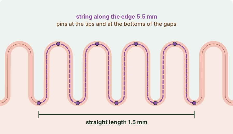
Supplement. A student lays a string along a stretch of the folded edge of a section of ileum, from the bottom of one gap to the bottom of another, and measures a straight line between the same two ends.
 (enlarge)
How many times longer is the folded edge than the straight length? What does this show, and what does it not show?
Times longer = string length ÷ straight length = 5.5 mm ÷ 1.5 mm = 3.7. Along this stretch the folded edge is about 3.7 times longer than a flat lining would be, so the villi increase the internal surface area of the small intestine. It does not give the real increase in surface area, which is larger, because the villi are fingers that add surface all round; and it does not measure a rate of absorption.
Microvilli: folds on every cell
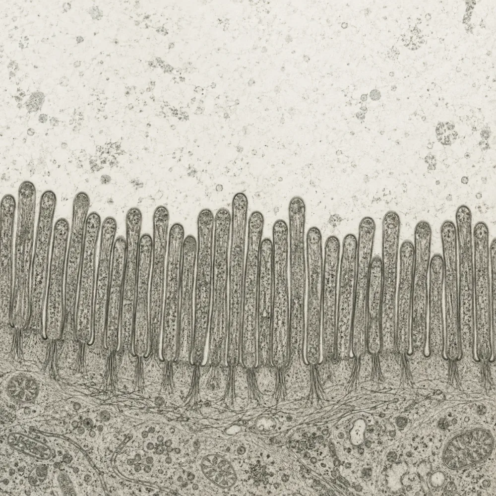
The villi are covered by a lining of cells, the epithelium. Each of its cells has a fringe of thousands of tiny projections on the side facing the food: these are microvilli. They are far too small to see one by one with a light microscope; at ×400 they show only as a fuzzy pink edge on the lining cells. An electron microscope shows them clearly.
 (enlarge)
So the lining is folded three times over: into ridges, the ridges into villi, and the surface of every lining cell into microvilli. A smooth tube 6 m long and 2.5 cm across would have an inner surface of about 0.5 m². The real lining of the small intestine has tens of square metres.
Trap: "Villi and microvilli are the same thing, just different sizes." A villus is a fold of the whole lining, made of many cells. Microvilli are part of a single cell: tiny projections of its membrane.
Worked example
Flattened villi
Supplement. In coeliac disease, eating gluten, a protein in wheat, damages the lining of the small intestine and the villi become short and flat. People with untreated coeliac disease are often tired and underweight, even when they eat enough. Explain why.
Flattened villi mean a smaller internal surface area of the small intestine. Absorption happens across this surface, so fewer nutrient molecules are absorbed in the time food takes to pass through. More glucose, amino acids, fatty acids, vitamins and minerals stay in the intestine and leave in the faeces. With less glucose absorbed for respiration, the person is tired; with fewer nutrients absorbed overall, they are underweight. On a gluten-free diet the villi grow back.
Key idea: (Supplement) The lining of the small intestine is covered with villi, and each cell of their epithelium has microvilli. Villi and microvilli greatly increase the internal surface area of the small intestine, so more nutrient molecules can be absorbed in the same time and absorption keeps pace with the food passing through.