Pupil sheet · The heart and double circulation
One beat, step by step — questions
Name ______________________ Date ____________
Try every question before you use a nudge. Write in full sentences where you are asked to explain.
1. identify
Supplement. The diagram shows a section through a mammal's heart. Which letter shows a semilunar valve?
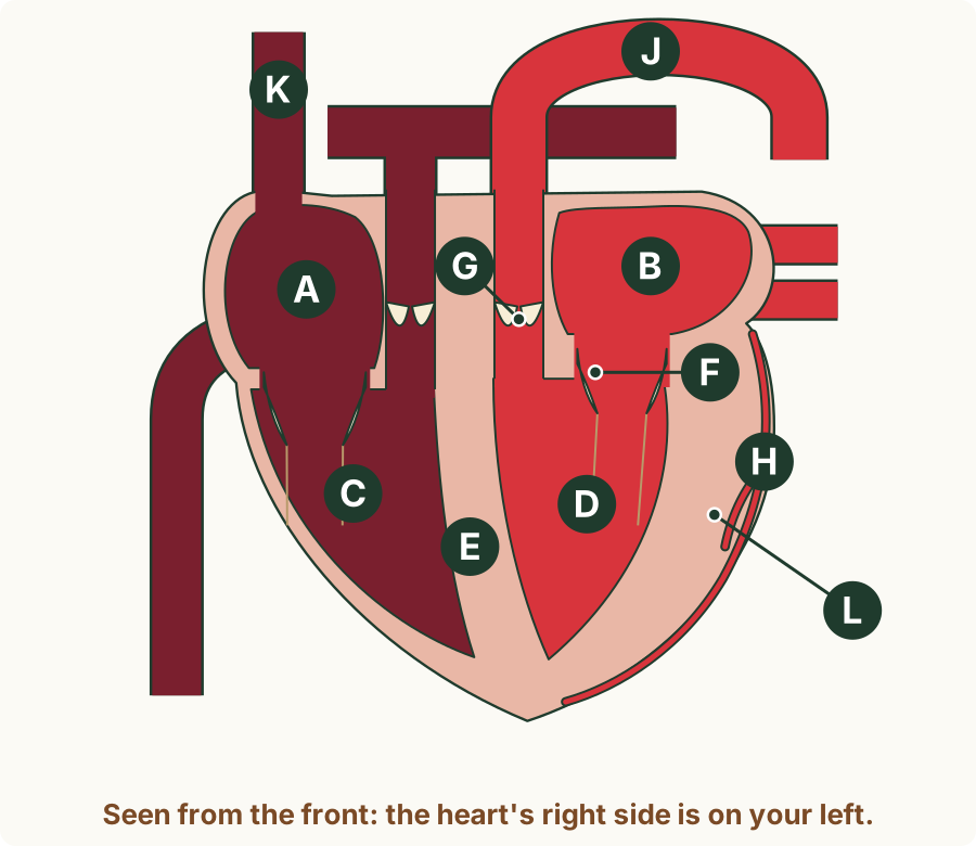
Circle one answer.
- H
- E
- G
- F
Nudge (only if you are stuck): There are two kinds of valve in the heart. Where does each kind sit?
2. sequence
Supplement. The ventricles have just started to contract, and the atrioventricular valves have just shut. Put in order what happens next, until the ventricles are ready to contract again.
Write the number of each step in order.
- the atria contract and squeeze their remaining blood into the ventricles
- the semilunar valves open and blood leaves the ventricles
- the ventricles relax and the semilunar valves shut
- the atrioventricular valves open and blood flows into the ventricles
- First: ______
- Second: ______
- Third: ______
- Last: ______
Nudge (only if you are stuck): A valve opens when the pressure behind it is higher than the pressure in front of it. Follow the pressure in the ventricles.
3. state
Supplement. Which part of the heart has the thickest muscle wall?
Circle one answer.
- the left atrium
- the right ventricle
- the right atrium
- the left ventricle
Nudge (only if you are stuck): Which part pumps blood the furthest?
4. state
Supplement. What does the septum keep apart?
Circle one answer.
- the blood inside the heart and the muscle of the heart's own wall
- the blood in the atria above and the blood in the ventricles below
- the arteries that leave the heart and the veins that come back to it
- oxygenated blood on the left and deoxygenated blood on the right
Nudge (only if you are stuck): The septum runs down the middle of the heart. What is on each side of it?
5. identify
Supplement. The ventricles are contracting and blood is leaving the heart. Which row is correct?
| Row | Atrioventricular valves | Semilunar valves |
|---|---|---|
| A | shut | open |
| B | open | shut |
| C | shut | shut |
| D | open | open |
Circle one answer.
- A
- B
- C
- D
Nudge (only if you are stuck): Where is the pressure higher: in the ventricles, or in the atria and the arteries?
6. identify / explain
Supplement. The graph shows the pressures in the right atrium, the right ventricle and the pulmonary artery during one beat. (a) Identify the numbered point at which the semilunar valve at the foot of the pulmonary artery shuts, and explain how the graph shows this. (b) Between points 1 and 2, the pressure in the right ventricle rises steeply, but no blood enters or leaves it. Explain why, using the pressures on the graph.
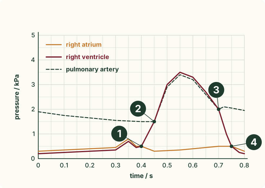
(a) The point, and how the graph shows it
(b) Between points 1 and 2
Nudge (only if you are stuck): For each pair of lines, ask which is higher at each moment, and which way that would push the blood.
7. explain
Supplement. In atrial fibrillation, the muscle of the atria only quivers and no longer contracts properly, yet many people live with it for years. In ventricular fibrillation, the ventricles only quiver, and the person dies within minutes unless treated. Explain, using this information, why the walls of the atria are thinner than the walls of the ventricles.
Nudge (only if you are stuck): What still happens to the blood when the atria only quiver, and what stops when the ventricles do?
8. identify
Supplement. The diagram shows a section through a sheep's heart, seen from the front, with four valves lettered. Which letter shows the atrioventricular valve on the heart's left side?
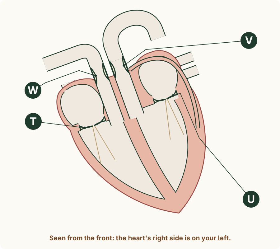
Circle one answer.
- U
- W
- V
- T
Nudge (only if you are stuck): First find the two valves that lie between two chambers. Then decide which side of the page is the heart's left side.
9. explain
Supplement. In a healthy person, the blood in the aorta carries as much oxygen as the blood in the pulmonary vein, and much more than the blood in the vena cava. Explain how the septum makes this possible, and why it matters to the body.
Nudge (only if you are stuck): Which side of the heart does each of the three vessels join?
10. identify
Supplement. Which row is correct for the valve at the foot of the pulmonary artery?
| Row | Kind of valve | It is shut when … | It stops blood flowing back into … |
|---|---|---|---|
| A | atrioventricular | the right ventricle relaxes | the right ventricle |
| B | semilunar | the right ventricle relaxes | the right ventricle |
| C | semilunar | the right ventricle contracts | the right atrium |
| D | atrioventricular | the right ventricle contracts | the right atrium |
Circle one answer.
- A
- B
- C
- D
Nudge (only if you are stuck): First decide the kind of valve from where it sits. Then ask when blood would try to flow back out of the artery.
11. identify / explain
Supplement. A student measures the thickness of the muscle wall of each of the four chambers of a dog's heart. (a) Identify which chamber, 1, 2, 3 or 4, is the left ventricle. (b) Explain how you know.
| Chamber | 1 | 2 | 3 | 4 |
|---|---|---|---|---|
| Wall thickness / mm | 4.5 | 1.6 | 11 | 1.9 |
(a) The left ventricle
(b) How you know
Nudge (only if you are stuck): Two walls are much thinner than the other two. Which pair are the atria?
12. suggest
Supplement. In some people, the semilunar valve at the foot of the aorta does not shut properly. Suggest what happens to some of the blood when the left ventricle relaxes, and how this affects the heart.
Nudge (only if you are stuck): After the ventricle relaxes, where is the pressure higher: in the aorta or in the ventricle?
13. suggest
Supplement. A frog's heart has two atria but only one ventricle. Oxygenated blood from the lungs and deoxygenated blood from the body both enter this one ventricle. Suggest a disadvantage of this heart compared with a mammal's.
Nudge (only if you are stuck): What does a mammal's septum do that the frog's single ventricle cannot?