Pupil sheet · Gas exchange and breathing
Lungs and gas exchange — questions
Name ______________________ Date ____________
Try every question before you use a nudge. Write in full sentences where you are asked to explain.
1. identify
The diagram shows the breathing system. Match each letter to the part.
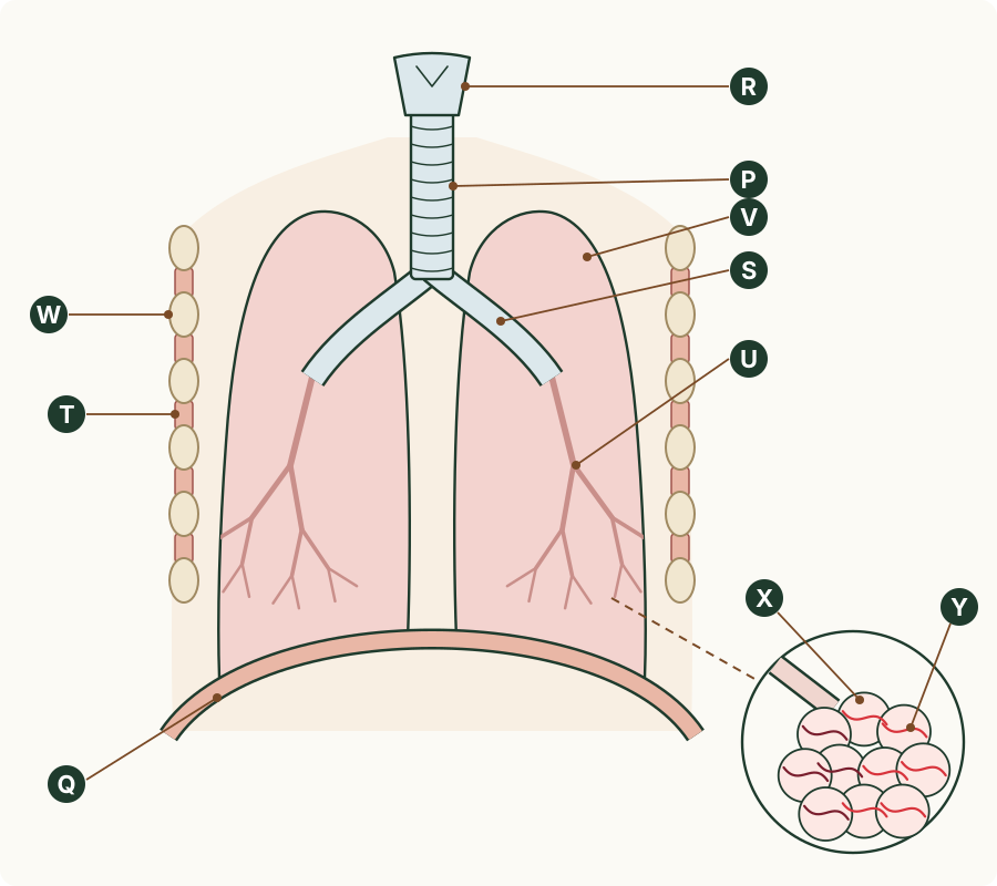
Write the number of the matching option.
- larynx
- bronchus
- lung
- diaphragm
- alveolus
- trachea
- intercostal muscle
- bronchiole
- capillary
- rib
- P: ______
- Q: ______
- R: ______
- S: ______
- T: ______
- U: ______
- V: ______
- W: ______
- X: ______
- Y: ______
Nudge (only if you are stuck): Follow the air from the neck down: the voice box, the windpipe, its branches, then the magnified circle.
2. describe
A doctor passes a thin tube with a camera on its tip (a bronchoscope) through a patient's mouth to look inside the right lung. Put the parts in the order the camera passes through them.
Write the number of each step in order.
- trachea
- a bronchiole
- larynx
- right bronchus
- First: ______
- Second: ______
- Third: ______
- Last: ______
Nudge (only if you are stuck): The camera goes down from the throat, and the tubes get narrower as it goes.
3. identify
In which part of the breathing system do oxygen and carbon dioxide pass between the air and the blood?
Circle one answer.
- the bronchi
- the trachea
- the alveoli
- the larynx
Nudge (only if you are stuck): Where does a breath end?
4. identify
Which features make the alveoli a good gas exchange surface? Choose all that apply.
Tick every correct statement.
- a few large air sacs
- good ventilation with air
- a thin surface
- thick walls
- a large surface area
- a good blood supply
Nudge (only if you are stuck): Each right answer helps diffusion.
5. explain
Match each feature of the alveoli to how it helps gas exchange.
Write the number of the matching option.
- the gases diffuse only a short distance
- the air in the alveoli is replaced again and again
- more gas can diffuse at the same time
- oxygen is carried away as soon as it enters the blood
- large surface area: ______
- thin surface: ______
- good blood supply: ______
- good ventilation with air: ______
Nudge (only if you are stuck): Which feature keeps the concentrations different on the two sides?
6. calculate / explain
One adult's alveoli have a total surface area of about 64 m². The same adult's skin has an area of about 1.7 m². (a) Calculate how many times larger the surface of the alveoli is than the skin. Give your answer to 2 significant figures. (b) The outer layer of the skin is many cells thick. Explain how the surface of the alveoli is different, and why this makes gas exchange faster.
(a) How many times larger: ______________ unit ________
(b) How the surface differs, and why it helps
Nudge (only if you are stuck): How many times larger = the larger area ÷ the smaller area.
7. explain
In one lung disease the thin walls between neighbouring alveoli break down, so the lungs have fewer, larger air sacs. Explain why the person becomes short of breath, especially when they exercise.
Nudge (only if you are stuck): Join many small sacs into a few large ones: what happens to the surface?
8. identify
Supplement. The diagram shows a section through the chest wall. Match each letter to the muscles it shows.
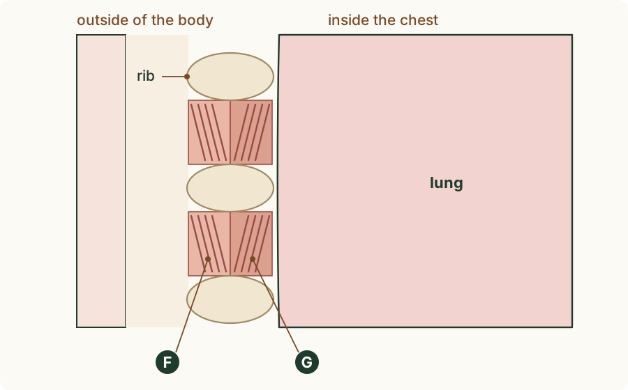
Write the number of the matching option.
- diaphragm
- external intercostal muscles
- internal intercostal muscles
- F: ______
- G: ______
Nudge (only if you are stuck): There are two layers between the ribs: which one is nearer the outside of the body?
9. describe / explain
Supplement. (a) Describe where the internal intercostal muscles are, compared with the external intercostal muscles. (b) Explain why the body needs two sets of intercostal muscles.
(a) Where the internal muscles are
(b) Why two sets
Nudge (only if you are stuck): A muscle can only pull.
10. state
Supplement. What is the function of the cartilage in the wall of the trachea?
Circle one answer.
- It traps dust and bacteria.
- It moves the ribs.
- It makes mucus.
- It keeps the trachea open.
Nudge (only if you are stuck): What would happen to a soft tube when the pressure inside it falls?
11. predict / explain
Supplement. Predict what would happen to the trachea during each breath in if its wall had no cartilage. Explain your answer.
Nudge (only if you are stuck): What happens to the pressure inside the airways when you breathe in?
12. identify
Supplement. The diagram shows the lining of the trachea. Match each letter to the part.
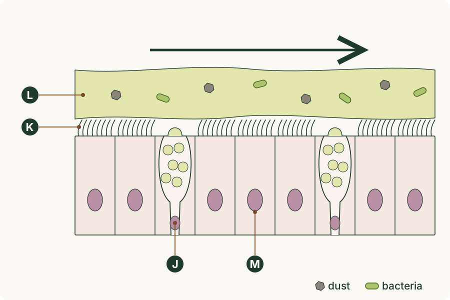
Write the number of the matching option.
- cilia
- mucus
- ciliated cell
- goblet cell
- J: ______
- K: ______
- L: ______
- M: ______
Nudge (only if you are stuck): Which layer is sticky, and which cells make it?
13. describe
Supplement. Complete the description of how the lining of the trachea protects the lungs. Goblet cells make (1), which traps (2) and dust. The (3) on ciliated cells beat and move it (4) the trachea to the throat.
(1)
(2)
(3)
(4)
Nudge (only if you are stuck): One gap is a direction.
14. state / explain
A child breathes in a small bead, which gets stuck in the right bronchus and blocks it. State which parts of the breathing system air can still reach, and explain why.
Nudge (only if you are stuck): Each bronchus leads to one lung.
15. suggest
The gills of a fish are made of many very thin filaments, each with a dense network of capillaries, and water flows over them all the time. Suggest how each of these features helps the fish take in oxygen.
Nudge (only if you are stuck): Use the same four features as the alveoli.
16. explain / identify
Supplement. (a) The biceps and the triceps in your arm are a pair of muscles that move the forearm in opposite directions. Explain how the external and internal intercostal muscles are like them. (b) A vacuum cleaner hose has a spiral of stiff wire in its wall. Identify the part of the breathing system that does the same job, and state the job.
(a) The intercostal muscles
(b) The part and its job
Nudge (only if you are stuck): Muscles pull; a stiff support stops a tube from flattening.
17. suggest
Supplement. Some people are born with cilia that cannot beat. Suggest why they get many chest infections.
Nudge (only if you are stuck): What normally moves the mucus out of the trachea?