Pupil sheet · The eye
The eye — final check
Name ______________________ Date ____________
Work alone, with the lesson closed. Answer every question; there are no hints.
1. identify
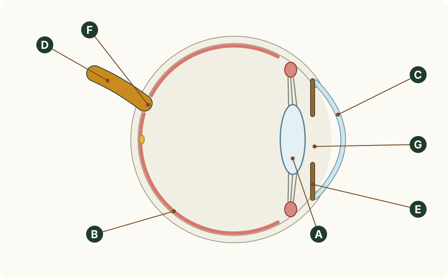
The diagram shows a cross-section of an eye drawn with the light arriving from the right. Identify the parts labelled A to G.
A
B
C
D
E
F
G
2. state
State the function of (a) the cornea, (b) the iris and (c) the optic nerve.
(a) Cornea
(b) Iris
(c) Optic nerve
3. identify / state
For each event, identify the sense organ that detects it and state the stimulus its receptors respond to: (a) you hear thunder; (b) you feel the warmth of the sun on your face.
(a) Thunder
(b) The sun's warmth
4. state / explain
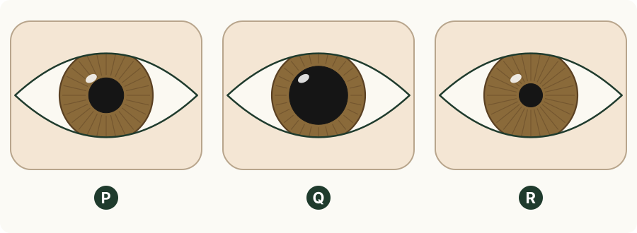
The drawings show the same person's eye, P, Q and R, in three different lights. (a) State the order of the eyes from the dimmest light to the brightest. (b) Explain how you decided.
(a) Dimmest to brightest
(b) How you decided
5. explain
There is a power cut at night, and a kitchen suddenly goes dark. Explain the pupil reflex that follows in the eyes of a person in the kitchen. Use the words stimulus, receptors, effector and response.
6. describe / explain
Supplement: describe how the muscles of the iris change the pupil when the light becomes bright, and explain why these muscles are described as antagonistic.
7. identify / state
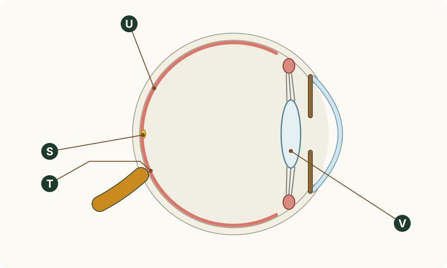
Supplement: the diagram shows a cross-section of an eye, with the light arriving from the right and four places lettered. (a) Identify the letter that shows the fovea. (b) State the function of the fovea. (c) Identify the letter that shows the place on the retina with no rods and no cones.
(a) Fovea
(b) Function
(c) No rods or cones
8. predict / explain
Supplement: after an injury, the ciliary muscles in one of a patient's eyes cannot contract. Predict whether that eye sees near objects or distant objects clearly, and explain your prediction.
9. explain
Supplement: a person has only two of the three kinds of cone. In bright daylight they cannot tell a red tomato from a green one, although they can see both tomatoes clearly. Explain this.
10. describe
Supplement: describe the distribution of rods and cones across the human retina.