Pupil sheet · Absorption
Inside a villus — questions
Name ______________________ Date ____________
Try every question before you use a nudge. Write in full sentences where you are asked to explain.
1. identify
Supplement. The diagram shows a villus cut along its length. Match each letter to the part it shows.
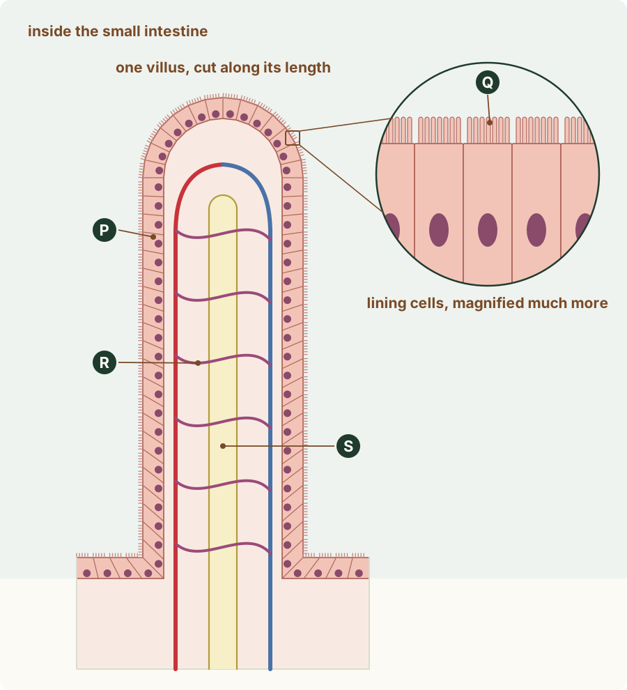
Write the number of the matching option.
- capillary network
- lacteal
- microvilli
- muscle layer
- epithelium
- P: ______
- Q: ______
- R: ______
- S: ______
Nudge (only if you are stuck): Start with the layer covering the surface, then the tube in the middle.
2. identify
Supplement. Which describes the layer that covers the surface of a villus?
Circle one answer.
- a thin layer of muscle
- an epithelium two cells thick
- an epithelium one cell thick
- an epithelium many cells thick
Nudge (only if you are stuck): At ×400 you saw the lining of the villus. Count its rows of cells.
3. state
Supplement. Complete the sentences about the vessels in a villus.
Glucose and amino acids are absorbed into the … .
Fatty acids and glycerol are absorbed into the … ,
which carries them away in the … .
Nudge (only if you are stuck): Two kinds of vessel; one carries blood and one carries lymph.
4. identify
Supplement. Put in order the places a fatty acid passes through, from the space inside the small intestine until it reaches the blood.
Write the number of each step in order.
- the lacteal in the middle of the villus
- the blood near the heart
- an epithelial cell of the villus
- larger lymph vessels outside the villus
- First: ______
- Second: ______
- Third: ______
- Last: ______
Nudge (only if you are stuck): Start at the surface facing the food. Then think which vessel in a villus takes the products of fat digestion, and where its fluid goes.
5. describe
Supplement. Describe the structure of a villus: (a) its shape and where it is; (b) its covering; (c) what is inside it.
(a) Shape and place
(b) Covering
(c) Inside
Nudge (only if you are stuck): Use the drawing of a villus cut along its length.
6. explain
Supplement. Explain how two features of the structure of a villus help absorption.
(a) First feature and how it helps
(b) Second feature and how it helps
Nudge (only if you are stuck): Think of distance, surface and what carries molecules away.
7. identify / suggest / explain
Supplement. Use the diagram. (a) Identify the fluid that flows through R and the fluid that flows through S. (b) S is closed at its tip. Suggest which way the fluid in S flows, and why. (c) Explain why it matters that blood flows through R all the time.
(a) The fluids in R and S
(b) Which way S flows, and why
(c) Why the flow in R matters
Nudge (only if you are stuck): R is a network of narrow vessels; S is the tube in the middle. For (b): where can fluid leave S?
8. predict
Supplement. A small blood clot blocks the arteriole that supplies one villus, so blood stops flowing through its capillaries. The rest of the gut is healthy. Which statement describes what happens to the absorption of glucose into that villus?
Circle one answer.
- It carries on as before, because the glucose now passes into the lacteal instead.
- It does not change, because the villus still has its epithelium and microvilli.
- It slows down, because glucose builds up in the still blood and is not carried away.
- It speeds up, because the glucose has more time to cross into the still blood.
Nudge (only if you are stuck): What does flowing blood do with glucose that has just been absorbed?
9. suggest / explain
Supplement. A weight-loss medicine stops lipase from working in the small intestine. Suggest how this changes what passes into the lacteals, and what happens to the fat in the meal.
(a) The change in the lacteals
(b) What happens to the fat
Nudge (only if you are stuck): What does lipase make, and where do those products go?
10. suggest / explain
Supplement. In one disease, the epithelium covering the villi becomes several cells thick. Suggest the effect on absorption, and explain why.
(a) The effect
(b) Why
Nudge (only if you are stuck): What must an absorbed molecule cross to reach the blood?
11. explain
Supplement. Lymph taken from the small intestine after a fatty meal looks milky white. After a meal of bread and boiled egg white, with no fat, it is almost clear. Explain the difference.
(a) After the fatty meal
(b) After the meal with no fat
Nudge (only if you are stuck): Which products of digestion go into the lymph, and which into the blood?